"Digestive system" and "alimentary system" redirect here. For digestive systems of non-human animals, see Digestion § Digestive systems. The digestive system The digestive system is a group of organs working together to convert food into energy and basic nutrients to feed the entire body. Food passes through a long tube inside the body known as the alimentary canal or the gastrointestinal tract (GI tract). The alimentary canal is made up of the oral cavity, pharynx, esophagus, stomach, small intestines, and large intestines. In addition to the alimentary canal, there are several important accessory organs that help your body to digest food...
The human digestive system consists of the gastrointestinal tract plus the accessory organs of digestion (the tongue, salivary glands, pancreas, liver, andgallbladder). In this system, the process of digestion has many stages, the first of which starts in the mouth (oral cavity). Digestion involves the breakdown of food into smaller and smaller components which can be absorbed and assimilated into the body. The secretion of saliva helps to produce a bolus which can be swallowed to pass down the oesophagus and into the stomach.
Saliva also contains a catalytic enzyme called amylase which starts to act on food in the mouth. Another digestive enzyme called lingual lipase is secreted by some of the lingual papillae on the tongue and also from serous glands in the main salivary glands. Digestion is helped by the mastication of food by the teethand also by the muscular actions of peristalsis and segmentation contractions. Gastric juice in the stomach is essential for the continuation of digestion as is the production of mucus in the stomach
Digestive system components

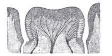
Historical depiction of the digestive system, 17th century Persia There are several organs and other components involved in the digestion of food. The organs known as the accessory digestive glands are the liver, gall bladderand pancreas. Other components include the mouth, teeth and epiglottis.
The largest structure of the digestive system is the gastrointestinal tract (GI tract). This starts at the mouth and ends at the anus, covering a distance of about nine (9) metres.[1]
The largest part of the GI tract is the colon or large intestine. Water is absorbed here and remaining waste matter is stored prior to defecation.[2]
Most of the digestion of food takes place in the small intestine.
A major digestive organ is the stomach. Within its mucosa are millions of embedded gastric glands. Their secretions are vital to the functioning of the organ.
There are many specialised cells of the GI tract. These include the various cells of the gastric glands, taste cells, pancreatic duct cells, enterocytes and microfold cells.
Mouth
The mouth is the first part of the gastrointestinal tract and is equipped with several structures that begin the first processes of digestion.[3] These include salivary glands, teeth and the tongue. The mouth consists of two regions, the vestibule and the oral cavity proper. The vestibule is the area between the teeth, lips and cheeks,[4] and the rest is the oral cavity proper. Most of the oral cavity is lined withoral mucosa, a mucous membrane that produces a lubricating mucus, of which only a small amount is needed. Mucous membranes vary in structure in the different regions of the body but they all produce a lubricating mucus, which is either secreted by surface cells or more usually by underlying glands. The mucous membrane in the mouth continues as the thin mucosa which lines the bases of the teeth. The main component of mucus is a glycoprotein called mucin and the type secreted varies according to the region involved. Mucin is viscous, clear, and clinging. Underlying the mucous membrane in the mouth is a thin layer of smooth muscle tissue and the loose connection to the membrane gives it its great elasticity.[5] It covers the cheeks, inner surfaces of the lips, and floor of the mouth.[6]
The roof of the mouth is termed the palate and it separates the oral cavity from the nasal cavity. The palate is hard at the front of the mouth since the overlying mucosa is covering a plate of bone; it is softer and more pliable at the back being made of muscle and connective tissue, and it can move to swallow food and liquids. The soft palate ends at the uvula.[7] The surface of the hard palate allows for the pressure needed in eating food, to leave the nasal passage clear.[8] The lips are the mouth's front boundary and the fauces (the passageway between the tonsils, also called the throat), mark its posterior boundary. At either side of the soft palate are the palatoglossus muscles which also reach into regions of the tongue. These muscles raise the back of the tongue and also close both sides of the fauces to enable food to be swallowed.[9] Mucus helps in the mastication of food in its ability to soften and collect the food in the formation of the bolus.
Salivary glands
There are three pairs of main salivary glands and between 800 and 1,000 minor salivary glands, all of which mainly serve the digestive process, and also play an important role in the maintenance of dental health and general mouth lubrication, without which speech would be impossible.[10] The main glands are all exocrine glands, secreting via ducts. All of these glands terminate in the mouth. The largest of these are the parotid glands – their secretion is mainly serous. The next pair are underneath the jaw, the submandibular glands, these produce both serous fluid and mucus. The serous fluid is produced by serous glands in these salivary glands which also produce lingual lipase. They produce about 70% of the oral cavity saliva. The third pair are the sublingual glands located underneath the tongue and their secretion is mainly mucous with a small percentage of saliva.
Within the oral mucosa (a mucous membrane) lining the mouth and also on the tongue and palates and mouth floor, are the minor salivary glands; their secretions are mainly mucous and are innervated by the facial nerve (the seventh cranial nerve).[11] The glands also secrete amylase a first stage in the breakdown of food acting on the carbohydrate in the food to transform the starch content into maltose. There are other glands on the surface of the tongue that encircle taste buds on the back part of the tongue and these also produce lingual lipase. Lipase is a digestive enzyme that catalyses the hydrolysis of lipids (fats). These glands are termed Von Ebner's glands which have also been shown to have another function in the secretion of histatins which offer an early defense (outside of the immune system) against microbes in food, when it makes contact with these glands on the tongue tissue.[10][12] Sensory information can stimulate the secretion of saliva providing the necessary fluid for the tongue to work with and also to ease swallowing of the food.
Saliva
Saliva functions initially in the digestive system to moisten and soften food into the formation of a bolus. The bolus is further helped by the lubrication provided by the saliva in its passage from the mouth into the oesophagus. Also of importance is the presence in saliva of the digestive enzymes amylase and lipase. Amylase starts to work on the starch in carbohydrates, breaking it down into the simple sugars of maltose and dextrose that can be further broken down in the small intestine. Saliva in the mouth can account for 30% of this initial starch digestion. Lipase starts to work on breaking downfats. Lipase is further produced in the pancreas where it is released to continue this digestion of fats. The presence of salivary lipase is of prime importance in young babies whose pancreatic lipase has yet to be developed.[13] As well as its role in supplying digestive enzymes, saliva has a cleansing action for the teeth and mouth.[14] It also has an immunological role in supplying antibodies to the system, such asimmunoglobulin A.[15] This is seen to be key in preventing infections of the salivary glands, importantly that of parotitis.
Saliva also contains a glycoprotein called haptocorrin which is a binding protein to vitamin B12.[16] It binds with the vitamin in order to carry it safely through the acidic content of the stomach. When it reaches the duodenum, pancreatic enzymes break down the glycoprotein and free the vitamin which then binds withintrinsic factor.
Tongue
Food enters the mouth where the first stage in the digestive process takes place, with the action of the tongue and the secretion of saliva. The tongue is a fleshy and muscular sensory organ, and the very first sensory information is received via the taste buds on its surface. If the taste is agreeable the tongue will go into action, manipulating the food in the mouth which stimulates the secretion of saliva from the salivary glands. The liquid quality of the saliva will help in the softening of the food and its enzyme content will start to break down the food whilst it is still in the mouth. The first part of the food to be broken down is the starch of carbohydrates. The tongue is attached to the floor of the mouth by a ligamentous band called the frenum[17] and this gives it great mobility for the manipulation of food (and speech); the range of manipulation is optimally controlled by the action of several muscles and limited in its external range by the stretch of the frenum. The tongue's two sets of muscles, are four intrinsic muscles that originate in the tongue and are involved with its shaping, and four extrinsic muscles originating in bone that are involved with its movement.
Taste
Taste is a form of chemoreception that takes place in the specialised receptors of taste cells, contained in structures called taste buds in the mouth. Taste buds are mainly on the upper surface (dorsum) of the tongue. Taste perception is vital to help prevent harmful or rotten foods from being consumed. This is a function of thegustatory system where the taste buds are at the forefront. There are taste buds elsewhere in the mouth not just on the surface of the tongue. The taste buds are innervated by a branch of the facial nerve the chorda tympani, and the glossopharyngeal nerve. Taste messages are sent via these cranial nerves to the brain. The brain can distinguish between the chemical qualities of the food. The five basic tastes are referred to as those of saltiness, sourness, bitterness and sweetness, and the most recent addition of a certain savouriness termed umami. The detection of saltiness and sourness enables the control of salt and acid balance. The detection of bitterness warns of poisons – many of a plant's defences are of poisonous compounds that are bitter. Sweetness guides to those foods that will supply energy; the initial breakdown of the energy-giving carbohydrates by salivary amylase creates the taste of sweetness since simple sugars are the first result. The taste of umami is thought to signal protein-rich food. Sour tastes are acidic which is often found in bad food. The brain has to decide very quickly whether to eat the food or not. It was the findings in 1991, describing the first olfactory receptors that helped to prompt the research into taste. The olfactory receptors are located on cell surfaces in the nose which bind to chemicals enabling the detection of smells. It is assumed that signals from taste receptors work together with the signals from those in the nose, to form an idea of complex food flavours.[18]
Teeth
Teeth are complex structures made of materials specific to them. They are made of a bone-like material called dentin, which is covered by the hardest tissue in the body—enamel.[19] Teeth have different shapes to deal with different aspects of mastication employed in tearing and chewing pieces of food into smaller and smaller pieces. This results in a much larger surface area for the action of digestive enzymes. The teeth are named after their particular roles in the process of mastication—incisors are used for cutting or biting off pieces of food; canines, are used for tearing, premolars and molars are used for chewing and grinding. Mastication of the food with the help of saliva and mucus results in the formation of a soft bolus which can then be swallowed to make its way down the upper gastrointestinal tract to the stomach.[20] The digestive enzymes in saliva also help in keeping the teeth clean by breaking down any lodged food particles. Epiglottis
The epiglottis is a flap that is made of elastic cartilage and attached to the entrance of the larynx. It is covered with a mucous membrane and there are taste buds on its lingual surface which faces into the mouth.[21] Its laryngeal surface faces into the larynx. The epiglottis functions to guard the entrance of the glottis, the opening between the vocal folds. It is normally pointed upward during breathing with its underside functioning as part of the pharynx, but during swallowing, the epiglottis folds down to a more horizontal position, with its upper side functioning as part of the pharynx. In this manner it prevents food from going into the trachea and instead directs it to the oesophagus, which is posterior. During swallowing, the backward motion of the tongue forces the epiglottis over the glottis' opening to prevent any food that is being swallowed from entering the larynx which leads to the lungs; the larynx is also pulled upwards to assist this process. Stimulation of the larynx by ingested matter produces a strong cough reflex in order to protect the lungs.
Pharynx
The pharynx is a part of the conducting zone of the respiratory system and also a part of the digestive system. It is the part of the throat immediately behind thenasal cavity at the back of the mouth and above the oesophagus and larynx. The pharynx is made up of three parts. The lower two parts–the oropharynx and thelaryngopharynx are involved in the digestive system. The laryngopharynx connects to the oesophagus and it serves as a passageway for both air and food. Air enters the larynx anteriorly but anything swallowed has priority and the passage of air is temporarily blocked. The pharynx is innervated by the pharyngeal plexus of the vagus nerve.[22] Muscles in the pharynx push the food into the oesophagus. The pharynx joins the oesophagus at the oesophageal inlet which is located behind thecricoid cartilage. Oesophagus
The oesophagus commonly known as the gullet, is an organ which consists of a muscular tube through which food passes from the pharynx to the stomach. The oesophagus is continuous with the laryngeal part of the pharynx. It passes through the posterior mediastinum in the thorax and enters the stomach through a hole in the thoracic diaphragm–the oesophageal hiatus, at the level of the tenththoracic vertebra (T10). Its length averages 25 cm, varying with height. It is divided into cervical, thoracic and abdominal parts. The pharynx joins the oesophagus at the oesophageal inlet which is behind the cricoid cartilage. At rest the oesophagus is closed at both ends, by the upper and lower oesophageal sphincters. The opening of the upper sphincter is triggered by the swallowing reflex so that food is allowed through. The sphincter also serves to prevent back flow from the oesophagus into the pharynx. The oesophagus has a mucous membrane and the epithelium which has a protective function is continuously replaced due to the volume of food that passes inside the oesophagus. During swallowing, food passes from the mouth through the pharynx into the oesophagus. The epiglottis folds down to a more horizontal position so as to prevent food from going into the trachea, instead directing it to the oesophagus. Once in the oesophagus, the bolus travels down to the stomach via rhythmic contraction and relaxation of muscles known as peristalsis. The lower oesophageal sphincter is a muscular sphincter surrounding the lower part of the oesophagus. The junction between the oesophagus and the stomach (the gastroesophageal junction) is controlled by the lower oesophageal sphincter, which remains constricted at all times other than during swallowing and vomiting to prevent the contents of the stomach from entering the oesophagus. As the oesophagus does not have the same protection from acid as the stomach, any failure of this sphincter can lead to heartburn. The oesophagus has a mucous membrane of epithelium which has a protective function as well as providing a smooth surface for the passage of food. Due to the high volume of food that is passed over time, this membrane is continuously renewed.
Diaphragm
The diaphragm is an important part of the body's digestive system. The diaphragm separates the thoracic cavity from the abdominal cavity where most of the digestive organs are located. The suspensory muscle attaches the ascending duodenum to the diaphragm. This muscle is thought to be of help in the digestive system in that its attachment offers a wider angle to the duodenojejunal flexure for the easier passage of digesting material. The diaphragm also attaches to thebare area of the liver, which it anchors. The oesophagus enters the abdomen through a hole in the diaphragm at the level of T10.
Stomach

Gastric acid (informally gastric juice), produced in the stomach plays a vital role in the digestive process, it mainly containshydrochloric acid and sodium chloride. A peptide hormone gastrin produced by G cells in the gastric glands, stimulates the production of gastric juice which activates the digestive enzymes. Pepsinogen is a precursor enzyme (zymogen) produced by the gastric chief cells and gastric acid activates this to the enzyme pepsin which begins the digestion of proteins. As these two chemicals would damage the stomach wall, mucus is secreted by innumerable gastric glands in the stomach, to provide a slimy protective layer against the damaging effects of the chemicals. At the same time that protein is being digested, mechanical churning occurs through the action of peristalsis, waves of muscular contractions that move along the stomach wall. This allows the mass of food to further mix with the digestive enzymes. Gastric lipase secreted by the chief cells in the fundic glands in the gastric mucosa of the stomach, is an acidic lipase, in contrast with the alkaline pancreatic lipase. This breaks down fats to some degree though is not as efficient as the pancreatic lipase.
The pylorus, the lowest section of the stomach which attaches to the duodenum via the pyloric canal, contains countless glands which secrete digestive enzymes including gastrin. After an hour or two, a thick semi-liquid called chyme is produced. When the pyloric sphincter, or valve opens, chyme enters the duodenum where it mixes further with digestive enzymes from the pancreas, and then passes through the small intestine, where digestion continues. When the chyme is fully digested, it is absorbed into the blood. 95% of absorption of nutrients occurs in the small intestine. Water and minerals are reabsorbed back into the blood in the colon of the large intestine, where the environment is slightly acidic. Some vitamins, such as biotin and vitamin K produced by bacteria in the gut flora of the colon are also absorbed.
The parietal cells in the fundus of the stomach, produce a glycoprotein called intrinsic factor which is essential for the absorption of vitamin B12. Vitamin B12 (cobalamin), is carried to, and through the stomach, bound to a glycoprotein secreted by the salivary glands - transcobalamin I also called haptocorrin, which protects the acid-sensitive vitamin from the acidic stomach contents. Once in the more neutral duodenum, pancreatic enzymes break down the protective glycoprotein. The freed vitamin B12 then binds to intrinsic factor which is then absorbed by the enterocytes in the ileum.
The stomach is a distensible organ and can normally expand to hold about one litre of food.[23] The stomach of a newborn baby will only be able to expand to retain about 30 ml.
Spleen
The spleen breaks down both red and white blood cells that are spent. This is why it is sometimes known as the 'graveyard of red blood cells'. A product of this digestion is the pigment bilirubin, which is sent to the liver and secreted in the bile. Another product is iron, which is used in the formation of new blood cells in the bone marrow.[5] Western medicine treats the spleen solely as belonging to thelymphatic system, though it is acknowledged that the full range of its important functions is not yet understood.[24] In contrast to this view, traditional Chinese medicine sees the spleen to be of central importance in the digestive system. The role of the spleen is seen to affect the health and vitality of the body in its turning of digested material from the stomach into usable nutrients and energy. Symptoms that include poor appetite, indigestion, bloating and jaundice, are seen to be indications of an imbalance in the spleen. The spleen is further seen to play a part in the metabolism of water, in ridding the body of excess fluid.[25] In the west, the spleen is seen to be paired with the stomach but in Chinese medicine, reference is made to the spleen system, which involves the pancreas. Fluids in the body are seen in traditional Chinese medicine to be under the control of the spleen. Fluids include digestive enzymes, saliva, mucus, fluid in the joints, tears, sweat and urine. They are categorised as thin and thick and together they are seen as nourishing all tissues and organs. In acupuncture two widely used acupuncture points - the stomach, (close to the knee) and the spleen, (halfway down from the knee) have long been seen to be connected and involved in digestive issues. Liver

The liver is the second largest organ (after the skin) and is an accessory digestive gland which plays a role in the body's metabolism. The liver has many functions some of which are important to digestion. The liver can detoxify various metabolites; synthesise proteins and produce biochemicals needed for digestion. It regulates the storage of glycogen which it can form from glucose (glycogenesis). The liver can also synthesise glucose from certain amino acids. Its digestive functions are largely involved with the breaking down of carbohydrates. It also maintains protein metabolism in its synthesis and degradation. In lipid metabolism it synthesises cholesterol. Fats are also produced in the process of lipogenesis. The liver synthesises the bulk of lipoproteins. The liver is located in the upper right quadrant of the abdomen and below the diaphragm to which it is attached at one part, This is to the right of the stomach and it overlies the gall bladder. The liver produces bile, an important alkaline compound which aids digestion.
Bile
Bile produced by the liver is made up of water (97%), bile salts, mucus and pigments, 1% fats and inorganic salts.[26] Bilirubin is its major pigment. Bile acts partly as a surfactant which lowers the surface tension between either two liquids or a solid and a liquid and helps to emulsify the fats in the chyme. Food fat is dispersed by the action of bile into smaller units called micelles. The breaking down into micelles creates a much larger surface area for the pancreatic enzyme, lipase to work on. Lipase digests the triglycerides which are broken down into two fatty acids and a monoglyceride. These are then absorbed by villi on the intestinal wall. If fats are not absorbed in this way in the small intestine problems can arise later in the large intestine which is not equipped to absorb fats. Bile also helps in the absorption of vitamin K from the diet. Bile is collected and delivered through the common hepatic duct. This duct joins with the cystic duct to connect in a common bile duct with the gallbladder. Bile is stored in the gallbladder for release when food is discharged into the duodenum and also after a few hours.[27]
Gallbladder
The gallbladder is a hollow part of the biliary system that sits just beneath the liver, with the gallbladder body resting in a small depression.[28] It is a small organ where the bile produced by the liver is stored, before being released into the small intestine. Bile flows from the liver through the bile ducts and into the gall bladder for storage. The bile is released in response to cholecystokinin (CKK) a peptide hormone released from the duodenum. The production of CKK (by endocrine cells of the duodenum) is stimulated by the presence of fat in the duodenum.[29]
It is divided into three sections, a fundus, body and neck. The neck tapers and connects to the biliary tree via the cystic duct, which then joins the common hepatic duct to form the common bile duct. At this junction is a mucosal fold called Hartmann's pouch, where gallstones commonly get stuck. The muscular layer of the body is of smooth muscle tissue that helps the gallbladder contract, so that it can discharge its bile into the bile duct. The gallbladder needs to store bile in a natural, semi-liquid form at all times. Hydrogen ions secreted from the inner lining of the gallbladder keep the bile acidic enough to prevent hardening. To dilute the bile, water and electrolytes from the digestion system are added. Also, salts attach themselves to cholesterol molecules in the bile to keep them from crystallising. If there is too much cholesterol or bilirubin in the bile, or if the gallbladder doesn't empty properly the systems can fail. This is how gallstones form when a small piece of calcium gets coated with either cholesterol or bilirubin and the bile crystallises and forms a gallstone. The main purpose of the gallbladder is to store and release bile, or gall. Bile is released into the small intestine in order to help in the digestion of fats by breaking down larger molecules into smaller ones. After the fat is absorbed, the bile is also absorbed and transported back to the liver for reuse.
Pancreas
Action of digestive hormones 
Pancreas, duodenum and bile duct The pancreas is a major organ functioning as an accessory digestive gland in the digestive system. It is both an endocrine gland and an exocrine gland.[30] The endocrine part secretes insulin when the blood sugar becomes high; insulin moves glucose from the blood into the muscles and other tissues for use as energy. The endocrine part releases glucagon when the blood sugar is low; glucagon allows stored sugar to be broken down into glucose by the liver in order to re–balance the sugar levels. The pancreas produces and releases important digestive enzymes in the pancreatic juice that it delivers to the duodenum. The pancreas lies below and at the back of the stomach. It connects to the duodenum via the pancreatic ductwhich it joins near to the bile duct's connection where both the bile and pancreatic juice can act on the chyme that is released from the stomach into the duodenum. Aqueous pancreatic secretions from pancreatic duct cells contain bicarbonate ions which are alkaline and help with the bile to neutralise the acidic chyme that is churned out by the stomach.
The pancreas is also the main source of enzymes for the digestion of fats and proteins. Some of these are released in response to the production of CKK in the duodenum. (The enzymes that digest polysaccharides, by contrast, are primarily produced by the walls of the intestines.) The cells are filled with secretory granules containing the precursor digestive enzymes. The major proteases, the pancreatic enzymes which work on proteins, are trypsinogen andchymotrypsinogen. Elastase is also produced. Smaller amounts of lipase and amylase are secreted. The pancreas also secretes phospholipase A2, lysophospholipase, and cholesterol esterase. The precursor zymogens, are inactive variants of the enzymes; which avoids the onset of pancreatitis caused by autodegradation. Once released in the intestine, the enzyme enteropeptidase present in the intestinal mucosa activates trypsinogen by cleaving it to form trypsin; further cleavage results in chymotripsin.
Lower gastrointestinal tract
The lower gastrointestinal tract (GI), includes the small intestine and all of the large intestine.[31] The intestine is also called the bowel or the gut. The lower GI starts at the pyloric sphincter of the stomach and finishes at the anus. The small intestine is subdivided into the duodenum, the jejunum and the ileum. The caecum marks the division between the small and large intestine. The large intestine includes the rectum and anal canal.[32][33] Small intestine

Lower GI tract - 3) Small intestine; 5) Caecum; 6) Large intestine Food starts to arrive in the small intestine one hour after it is eaten, and after two hours the stomach has emptied. Until this time the food is termed a bolus. It then becomes the partially digested semi-liquid termed chyme. In the small intestine, the pH becomes crucial; it needs to be finely balanced in order to activate digestive enzymes. The chyme is very acidic, with a low pH, having been released from the stomach and needs to be made much more alkaline. This is achieved in the duodenum by the addition of bile from the gall bladder combined with thebicarbonate secretions from the pancreatic duct and also from secretions of bicarbonate-rich mucus from duodenal glands known as Brunner's glands. The chyme arrives in the intestines having been released from the stomach through the opening of the pyloric sphincter. The resulting alkaline fluid mix neutralises the gastric acid which would damage the lining of the intestine. The mucus component lubricates the walls of the intestine. When the digested food particles are reduced enough in size and composition, they can be absorbed by the intestinal wall and carried to the bloodstream. The first receptacle for this chyme is the duodenal bulb. From here it passes into the first of the three sections of the small intestine, the duodenum. (The next section is the jejunum and the third is theileum). The duodenum is the first and shortest section of the small intestine. It is a hollow, jointed C-shaped tube connecting the stomach to the jejunum. It starts at the duodenal bulb and ends at the suspensory muscle of duodenum. The attachment of the suspensory muscle to the diaphragm is thought to help the passage of food by making a wider angle at its attachment.
Most food digestion takes place in the small intestine. In the duodenum, pancreatic lipase is secreted together with a co-enzyme, colipase to further digest the fat content of the chyme. From this breakdown, smaller particles of emulsified fats called chylomicrons are produced. There are also digestive cells called enterocyteslining the intestines (the majority being in the small intestine). They are unusual cells in that they have villi on their surface which in turn have innumerable microvilli on their surface. All these villi make for a greater surface area, not only for the absorption of chyme but also for its further digestion by large numbers of digestive enzymes present on the microvilli.
The cholymicrons are small enough to pass through the enterocyte villi and into their lymph capillaries called lacteals. A milky fluid called chyle consisting mainly of the emulsified fats of the cholymicrons results from the absorbed mix with the lymph in the lacteals. Chyle is then transported through the lymphatic system to the rest of the body.
The suspensory muscle marks the end of the duodenum and the division between the upper gastrointestinal tract and the lower GI tract. The digestive tract continues as the jejunum which continues as the ileum. The jejunum, the midsection of the small intestine contains circular folds, flaps of doubled mucosal membrane which partially encircle and sometimes completely encircle the lumen of the intestine. These folds together with villi serve to increase the surface area of the jejunum enabling an increased absorption of digested sugars, amino acids and fatty acids into the bloodstream. The circular folds also slow the passage of food giving more time for nutrients to be absorbed.
The last part of the small intestine is the ileum. This also contains villi and vitamin B12; bile acids and any residue nutrients are absorbed here. When the chyme is exhausted of its nutrients the remaining waste material changes into the semi-solids called faeces, which pass to the large intestine, where bacteria in the gut flora further break down residual proteins and starches.[34]
Caecum

Caecum and beginning of ascending colon The caecum is a pouch marking the division between the small intestine and the large intestine.[35] The caecum receives chyme from the last part of the small intestine, the terminal ileum, and connects to the ascending colon of the large intestine. At this junction there is a sphincter or valve, the ileocecal valve which slows the passage of chyme from the ileum, allowing further digestion. It is also the site of the appendix attachment.
Large intestine
In the large intestine, the passage of the digesting food in the colon is a lot slower, taking from 12 to 50 hours until it is removed by defecation. The colon mainly serves as a site for the fermentation of digestible matter by the gut flora. The time taken varies considerably between individuals. The remaining semi-solid waste is termed faeces and is removed by the coordinated contractions of the intestinal walls, termed peristalsis, which propels the excreta forward to reach therectum and exit via defecation from the anus. The wall has an outer layer of longitudinal muscles, the taeniae coli, and an inner layer of circular muscles. The circular muscle keeps the material moving forward and also prevents any back flow of waste. Also of help in the action of peristalsis is the basal electrical rhythm that determines the frequency of contractions.[36] The taeniae coli can be seen and are responsible for the bulges (haustra) present in the colon. Most parts of the GI tract are covered with serous membranes and have a mesentery. Other more muscular parts are lined with adventitia.
Blood supply
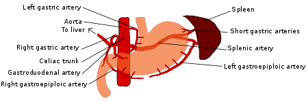
Blood supply to the digestive organs[37] 
Arteries and veins around the pancreas and spleen The digestive system is supplied by the celiac artery. The celiac artery is the first major branch from the abdominal aorta, and is the only major artery that nourishes the digestive organs.
There are three main divisions – the left gastric artery, the common hepatic artery and thesplenic artery.
The celiac artery supplies the liver, stomach, spleen and the upper 1/3 of the duodenum (to thesphincter of Oddi) and the pancreas with oxygenated blood. Most of the blood is returned to the liver via the portal venous system for further processing and detoxification before returning to the systemic circulation via the hepatic portal vein.
The next branch from the abdominal aorta is the superior mesenteric artery, which supplies the regions of the digestive tract derived from the midgut, which includes the distal 2/3 of the duodenum, jejunum, ileum, caecum, appendix, ascending colon, and the proximal 2/3 of the transverse colon.
The final branch which is important for the digestive system is the inferior mesenteric artery, which supplies the regions of the digestive tract derived from the hindgut, which includes the distal 1/3 of the transverse colon, descending colon, sigmoid colon, rectum, and the anus above the pectinate line.
Innervation
The enteric nervous system consists of some one hundred million neurons[38] that are embedded in the peritoneum, the lining of the gastrointestinal tractextending from the oesophagus to the anus.[39] These neurons are collected into two plexuses - the myenteric (or Auerbach's) plexus that lies between the longitudinal and the smooth muscle layers, and the submucosal (or Meissner's) plexus that lies between the circular smooth muscle layer and the mucosa.[40][41][42]
Parasympathetic innervation to the ascending colon is supplied by the vagus nerve. Sympathetic innervation is supplied by the splanchnic nerves that join theceliac ganglia. Most of the digestive tract is innervated by the two large celiac ganglia, with the upper part of each ganglion joined by the greater splanchnic nerve and the lower parts joined by the lesser splanchnic nerve. It is from these ganglia that many of the gastric plexuses arise.
Clinical significance
Each part of the digestive system is subject to a wide range of disorders. A number of problems including malnutrition and anemia can arise from malabsorption, the abnormal absorption of nutrients in the GI tract. Malabsorption can have many causes ranging from infection, to enzyme deficiencies such as exocrine pancreatic insufficiency. It can also arise as a result of other gastrointestinal diseases such as coeliac disease. Coeliac disease is an autoimmune disorder of the small intestine. This can cause vitamin deficiencies due to the improper absorption of nutrients in the small intestine. In the oesophagus Schatzki rings can restrict the passageway, causing difficulties in swallowing. They can also completely block the oesophagus.[43]
A common disorder of the bowel is diverticulitis. Diverticula are small pouches that can form inside the bowel wall, which can become inflamed to give diverticulitis. This disease can have complications if an inflamed diverticulum bursts and infection sets in. Any infection can spread further to the lining of the abdomen (peritoneum) and cause potentially fatal peritonitis.[44]
Crohn's disease is a common chronic inflammatory bowel disease (IBD), which can affect any part of the GI tract,[45] but it mostly starts in the terminal ileum.
Ulcerative colitis an ulcerative form of colitis, is the other major inflammatory bowel disease which is restricted to the colon and rectum. Both of these IBDs can give an increased risk of the development of colorectal cancer. Ulcerative coliltis is the most common of the IBDs[46]
Irritable bowel syndrome (IBS) is the most common of the functional gastrointestinal disorders. These are idiopathic disorders that the Rome process has helped to define.[47]
Giardiasis is a disease of the small intestine caused by a protist parasite Giardia lamblia. This does not spread but remains confined to the lumen of the small intestine.[48] It can often be asymptomatic, but as often can be indicated by a variety of symptoms. Giardiasis is the most common pathogenic parasitic infection in humans.[49]
There are diagnostic tools mostly involving the ingestion of barium sulphate to investigate disorders of the GI tract.[50] These are known as upper gastrointestinal series that enable imaging of the pharynx, larynx, oesophagous, stomach and small intestine and lower gastrointestinal series for imaging of the colon.
Mouth
The mouth is the beginning of the digestive tract. In fact, digestion starts here as soon as you take the first bite of a meal. Chewing breaks the food into pieces that are more easily digested, while saliva mixes with food to begin the process of breaking it down into a form your body can absorb and use.
Throat
Also called the pharynx, the throat is the next destination for food you've eaten. From here, food travels to the esophagus or swallowing tube.
Esophagus
The esophagus is a muscular tube extending from the pharynx to the stomach. By means of a series of contractions, called peristalsis, the esophagus delivers food to the stomach. Just before the connection to the stomach there is a "zone of high pressure," called the lower esophageal sphincter; this is a "valve" meant to keep food from passing backwards into the esophagus.
Stomach
The stomach is a sac-like organ with strong muscular walls. In addition to holding the food, it's also a mixer and grinder. The stomach secretes acid and powerful enzymes that continue the process of breaking down the food. When it leaves the stomach, food is the consistency of a liquid or paste. From there the food moves to the small intestine.
Small Intestine
Made up of three segments, the duodenum, jejunum, and ileum, the small intestine is a long tube loosely coiled in the abdomen (spread out, it would be more than 20 feet long). The small intestine continues the process of breaking down food by using enzymes released by the pancreas and bile from the liver. Bile is a compound that aids in the digestion of fat and eliminates waste products from the blood. Peristalsis (contractions) is also at work in this organ, moving food through and mixing it up with digestive secretions. The duodenum is largely responsible for continuing the process of breaking down food, with the jejunum and ileum being mainly responsible for the
Small Intestine continued...
Three organs play a pivotal role in helping the stomach and small intestine digest food:
Pancreas
Among other functions, the oblong pancreas secretes enzymes into the small intestine. These enzymes break down protein, fat, and carbohydrates from the food we eat.
Liver The liver has many functions, but two of its main functions within the digestive systemare to make and secrete bile, and to cleanse and purify the blood coming from the small intestine containing the nutrients just absorbed.
Gallbladder
The gallbladder is a pear-shaped reservoir that sits just under the liver and stores bile. Bile is made in the liver then travels to the gallbladder through a channel called the cystic duct. During a meal, the gallbladder contracts, sending bile to the small intestine.
Once the nutrients have been absorbed and the leftover liquid has passed through the small intestine, what is left of the food you ate is handed over to the large intestine, orcolon.
Colon (Large Intestine)
The colon is a 5- to 6-foot-long muscular tube that connects the cecum (the first part of the large intestine to the rectum (the last part of the large intestine). It is made up of the cecum, the ascending (right) colon, the transverse (across) colon, the descending (left) colon, and the sigmoid colon (so-called for its "S" shape; the Greek letter for S is called the sigma), which connects to the rectum.
Stool, or waste left over from the digestive process, is passed through the colon by means of peristalsis (contractions), first in a liquid state and ultimately in solid form as the water is removed from the stool. A stool is stored in the sigmoid colon until a "mass movement" empties it into the rectum once or twice a day. It normally takes about 36 hours for stool to get through the colon. The stool itself is mostly food debris and bacteria. These bacteria perform several useful functions, such as synthesizing various vitamins, processing waste products and food particles, and protecting against harmful bacteria. When the descending colon becomes full of stool, or feces, it empties its contents into the rectum to begin the process of elimination.
Rectum
The rectum (Latin for "straight") is an 8-inch chamber that connects the colon to the anus. It is the rectum's job to receive stool from the colon, to let you know there is stool to be evacuated, and to hold the stool until evacuation happens. When anything (gas or stool) comes into the rectum, sensors send a message to the brain. The brainthen decides if the rectal contents can be released or not. If they can, the sphincters (muscles) relax and the rectum contracts, expelling its contents. If the contents cannot be expelled, the sphincters contract and the rectum accommodates, so that the sensation temporarily goes away.
Anus
The anus is the last part of the digestive tract. It consists of the pelvic floor muscles and the two anal sphincters (internal and external muscles). The lining of the upper anus is specialized to detect rectal contents. It lets us know whether the contents are liquid, gas, or solid. The pelvic floor muscle creates an angle between the rectum and the anus that stops stool from coming out when it is not supposed to. The anal sphincters provide fine control of stool. The internal sphincter keeps us from going to the bathroom when we are asleep, or otherwise unaware of the presence of stool. When we get an urge to go to the bathroom, we rely on our external sphincter to keep the stool in until we can get to the toilet.
The digestive system is responsible for taking whole foods and turning them into energy and nutrients to allow the body to function, grow, and repair itself. The six primary processes of the digestive system include:
- Ingestion of food
- Secretion of fluids and digestive enzymes
- Mixing and movement of food and wastes through the body
- Digestion of food into smaller pieces
- Absorption of nutrients
- Excretion of wastes
Ingestion
The first function of the digestive system is ingestion, or the intake of food. The mouth is responsible for this function, as it is the orifice through which all food enters the body. The mouth and stomach are also responsible for the storage of food as it is waiting to be digested. This storage capacity allows the body to eat only a few times each day and to ingest more food than it can process at one time.
Secretion
In the course of a day, the digestive system secretes around 7 liters of fluids. These fluids include saliva, mucus, hydrochloric acid, enzymes, and bile. Saliva moistens dry food and contains salivary amylase, a digestive enzyme that begins the digestion of carbohydrates. Mucus serves as a protective barrier and lubricant inside of the GI tract. Hydrochloric acid helps to digest food chemically and protects the body by killing bacteria present in our food. Enzymes are like tiny biochemical machines that disassemble large macromolecules like proteins, carbohydrates, and lipids into their smaller components. Finally, bile is used to emulsify large masses of lipids into tiny globules for easy digestion.
Mixing and Movement
The digestive system uses 3 main processes to move and mix food:
- Swallowing. Swallowing is the process of using smooth and skeletal muscles in the mouth, tongue, and pharynx to push food out of the mouth, through the pharynx, and into the esophagus.
- Peristalsis. Peristalsis is a muscular wave that travels the length of the GI tract, moving partially digested food a short distance down the tract. It takes many waves of peristalsis for food to travel from the esophagus, through the stomach andintestines, and reach the end of the GI tract.
- Segmentation. Segmentation occurs only in the small intestine as short segments of intestine contract like hands squeezing a toothpaste tube. Segmentation helps to increase the absorption of nutrients by mixing food and increasing its contact with the walls of the intestine.
Digestion
Digestion is the process of turning large pieces of food into its component chemicals. Mechanical digestion is the physical breakdown of large pieces of food into smaller pieces. This mode of digestion begins with the chewing of food by the teeth and is continued through the muscular mixing of food by the stomach and intestines. Bile produced by the liver is also used to mechanically break fats into smaller globules. While food is being mechanically digested it is also being chemically digested as larger and more complex molecules are being broken down into smaller molecules that are easier to absorb. Chemical digestion begins in the mouth with salivary amylase in saliva splitting complex carbohydrates into simple carbohydrates. The enzymes and acid in the stomach continue chemical digestion, but the bulk of chemical digestion takes place in the small intestine thanks to the action of the pancreas. The pancreas secretes an incredibly strong digestive cocktail known as pancreatic juice, which is capable of digesting lipids, carbohydrates, proteins and nucleic acids. By the time food has left the duodenum, it has been reduced to its chemical building blocks—fatty acids, amino acids, monosaccharides, and nucleotides.
Absorption
Once food has been reduced to its building blocks, it is ready for the body to absorb. Absorption begins in the stomach with simple molecules like water and alcohol being absorbed directly into the bloodstream. Most absorption takes place in the walls of the small intestine, which are densely folded to maximize the surface area in contact with digested food. Small blood and lymphatic vessels in the intestinal wall pick up the molecules and carry them to the rest of the body. The large intestine is also involved in the absorption of water and vitamins B and K before feces leave the body.
Excretion
The final function of the digestive system is the excretion of waste in a process known as defecation. Defecation removes indigestible substances from the body so that they do not accumulate inside the gut. The timing of defecation is controlled voluntarily by the conscious part of the brain, but must be accomplished on a regular basis to prevent a backup of indigestible materials.
Digestive System: Facts, Function & Diseases
The human digestive system is a series of organs that converts food into essential nutrients that are absorbed into the body and eliminates unused waste material. It is essential to good health because if the digestive system shuts down, the body cannot be nourished or rid itself of waste.
Description of the digestive system
Also known as the gastrointestinal (GI) tract, the digestive system begins at the mouth, includes the esophagus, stomach, small intestine, large intestine (also known as the colon) and rectum, and ends at the anus. The entire system — from mouth to anus — is about 30 feet (9 meters) long, according to the American Society of Gastrointestinal Endoscopy (ASGE).
Digestion begins with the mouth. Even the smell of food can generate saliva, which is secreted by the salivary glands in the mouth, contains an enzyme, salivary amylase, which breaks down starch. Teeth, which are part of the skeletal system, play a key role in digestion. In carnivores, teeth are designed for killing and breaking down meat. Herbivores’ teeth are made for grinding plants and other food to ease them through the digestion process. Swallowing pushes chewed food into the esophagus, where it passes through the oropharynx and hypopharynx. At this point, food takes the form of a small round mass and digestion becomes involuntary. A series of muscular contractions, called peristalsis, transports food through the rest of the system. The esophagus empties into the stomach, according to the National Institutes of Health (NIH).
The stomach’s gastric juice, which is primarily a mix of hydrochloric acid and pepsin, starts breaking down proteins and killing potentially harmful bacteria, according to ASGE. After an hour or two of this process, a thick semi-liquid paste, called chyme, forms.
At this point the pyloric sphincter valve opens and chyme enters the duodenum, where it mixes with digestive enzymes from the pancreas and acidic bile from the gall bladder, according to the Cleveland Clinic. The next stop for the chyme is the small intestine, a 20-foot (6-meter) tube-shaped organ, where the majority of the absorption of nutrients occurs. The nutrients move into the bloodstream and are transported to the liver.
The liver creates glycogen from sugars and carbohydrates to give the body energy and converts dietary proteins into new proteins needed by the blood system. The liver also breaks down unwanted chemicals, such as alcohol, which is detoxified and passed from the body as waste, the Cleveland Clinic noted.
Whatever material is left goes into the large intestine. The function of the large intestine, which is about 5 feet long (1.5 meters), is primarily for storage and fermentation of indigestible matter. Also called the colon, it has four parts: the ascending colon, the transverse colon, the descending colon and the sigmoid colon. This is where water from the chyme is absorbed back into the body and feces are formed primarily from water (75 percent), dietary fiber and other waste products, according to the Cleveland Clinic. Feces are stored here until they are eliminated from the body through defecation.Swallowing pushes chewed food into the esophagus, where it passes through the oropharynx and hypopharynx. At this point, food takes the form of a small round mass and digestion becomes involuntary. A series of muscular contractions, called peristalsis, transports food through the rest of the system. The esophagus empties into the stomach, according to the National Institutes of Health (NIH).
The stomach’s gastric juice, which is primarily a mix of hydrochloric acid and pepsin, starts breaking down proteins and killing potentially harmful bacteria, according to ASGE. After an hour or two of this process, a thick semi-liquid paste, called chyme, forms.
At this point the pyloric sphincter valve opens and chyme enters the duodenum, where it mixes with digestive enzymes from the pancreas and acidic bile from the gall bladder, according to the Cleveland Clinic. The next stop for the chyme is the small intestine, a 20-foot (6-meter) tube-shaped organ, where the majority of the absorption of nutrients occurs. The nutrients move into the bloodstream and are transported to the liver.
The liver creates glycogen from sugars and carbohydrates to give the body energy and converts dietary proteins into new proteins needed by the blood system. The liver also breaks down unwanted chemicals, such as alcohol, which is detoxified and passed from the body as waste, the Cleveland Clinic noted.
Whatever material is left goes into the large intestine. The function of the large intestine, which is about 5 feet long (1.5 meters), is primarily for storage and fermentation of indigestible matter. Also called the colon, it has four parts: the ascending colon, the transverse colon, the descending colon and the sigmoid colon. This is where water from the chyme is absorbed back into the body and feces are formed primarily from water (75 percent), dietary fiber and other waste products, according to the Cleveland Clinic. Feces are stored here until they are eliminated from the body through defecation.
Diseases of the digestive system
Many symptoms can signal problems with the GI tract, including: abdominal pain, blood in the stool, bloating, constipation, diarrhea, heartburn, incontinence, nausea and vomiting and difficulty swallowing, according to the NIH.
Among the most widely known diseases of the digestive system is colon cancer. According to the Centers for Disease Control (CDC), 51,783 Americans died from colon cancer in 2011 (the most recent year for available data). Excluding skin cancers, colon and rectal cancer, or colorectal cancer, is the third most common cancer diagnosed in both men and women in the United States, according to the American Cancer Society.
Polyp growth and irregular cells, which may or may not be cancerous, are the most common development paths for colorectal cancers (also referred to as CRC), and can be detected during a routine colonoscopy, according to Dr. John Marks, a gastroenterologist affiliated with the Main Line Health health care system.
What are digestive problems?
Digestive symptoms include a wide variety of symptoms that affect the digestive or gastrointestinal system. The gastrointestinal system includes the throat, esophagus, stomach, small intestine, large intestine, anus, pancreas, liver, and gallbladder. Digestive symptoms can be due to a wide variety of mild to serious diseases, disorders and conditions. They can occur in all age groups and populations.
Common digestive symptoms include:
Digestive symptoms can vary greatly in character and severity depending on the underlying disease, disorder or condition. Depending on the cause, digestive symptoms can last briefly and disappear quickly, such as symptoms that occur during a single episode of indigestion. Digestive symptoms can also persist or recur over a longer period of time, such as when due to colorectal cancer or inflammatory bowel disease.
What other symptoms might occur with digestive symptoms?
Digestive symptoms may be accompanied by symptoms in other body systems depending on the underlying disease, disorder or condition. Other symptoms that may occur along with digestive symptoms include:
Chest pain or pressure
Chills
Dizziness
Easy bleeding or bruising
Flu-like symptoms (fatigue, fever, sore throat, headache, cough, aches and pains)
Jaundice (yellowing of skin and eyes)
Pale skin
Referred shoulder pain
Weakness (loss of strength)
Weight loss, malabsorption, and vitamin deficiencies
Serious symptoms that might indicate a life-threatening condition
In some cases, digestive symptoms may occur with other symptoms that might indicate a serious or life-threatening condition that should be immediately evaluated in an emergency setting. Seek immediate medical care (call 911) if you, or someone you are with, have any of these symptoms:
What causes digestive symptoms?
Digestive symptoms have many possible underling causes. General conditions that can cause digestive symptoms include infection, malignancy, inflammation, trauma, obstruction, and other abnormal processes.
Digestive symptoms can result from gastrointestinal or digestive conditions or from conditions of other body systems, such as the endocrine system, the nervous system, the reproductive system, and the urinary system.
Gastrointestinal causes of digestive symptoms
Appendicitis
Celiac disease (severe sensitivity to gluten from wheat and other grains that causes intestinal damage)
Colorectal cancer, gastric cancer, and pancreatic cancer
Diverticulitis and diverticulosis
Food poisoning
Gallstones
Gastroesophageal reflux disease (GERD)
Inflammatory bowel disease (includes Crohn’s disease and ulcerative colitis)
Intestinal obstruction
Irritable bowel syndrome (IBS; digestive discomfort that does not cause intestinal damage or serious disease)
Lactose intolerance and other food intolerances (fructose, gluten)
Lactose intolerance and other food intolerances
Liver disease (hepatitis, cirrhosis, liver failure)
Pancreatitis
Peptic ulcer
Viral gastroenteritis
Other causes of digestive symptoms
Other causes of digestive symptoms include:
Altitude sickness
Anxiety and depression
Chronic dehydration
Concussion
Exposure to smoke or toxic fumes or substances
Inadequate daily consumption of dietary fiber
Kidney disease (kidney stone, kidney failure, pyelonephritis)
Medication side effects (antibiotics, anticholinergics, opioids, many others)
Meniere’s disease
Migraine
Motion sickness
Narcotic use or withdrawal
Pregnancy and labor
Stress
Type 1 or type 2 diabetes
Vertigo
Life-threatening causes of digestive swelling
In some cases, digestive symptoms may accompany a serious or life-threatening condition that should be immediately evaluated in an emergency setting. These include
What are the potential complications of digestive symptoms?
In some cases, digestive symptoms can lead to serious complications, especially if the underlying disease or condition is untreated or poorly managed. Once the underlying cause is identified, you can help minimize your risk of serious complications by following the treatment plan you and your health care professional design specifically for you. Complications include:
Bowel obstruction
Dehydration due to vomiting, diarrhea, or a decreased desire to drink fluids
Difficulty breathing
Erosive esophagitis
Peritonitis
Poor nutrition due to vomiting, diarrhea, or a decreased desire to eat
Reduced appetite
Sepsis (life-threatening bacterial blood infection)
Severe discomfort or pain
Shock
.


.jpg)