The oral cavity includes the lips, the inside lining of the lips and cheeks (buccal mucosa), the teeth, the gums, the front two-thirds of the tongue, the floor of the mouth below the tongue, and the bony roof of the mouth (hard palate).Your dentist can usually detect tooth decay easily by:
Asking about tooth pain and sensitivity
Examining your mouth and teeth
Probing your teeth with dental instruments to check for soft areas
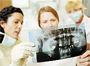
Looking at dental X-rays, which can show the extent of cavities and decay
Your dentist will also be able to tell you which of the three types of cavities you have — smooth surface, pit and fissure, or root.
In addition to its primary role as the beginning of the digestive system, in humans the mouth also plays a significant role incommunication. While primary aspects of thevoiceare produced in thethroat, thetongue,lips, andjaware also needed to produce the range of sounds included in human language.
The mouth consists of two regions, thevestibuleand theoral cavity proper. The mouth, normally moist, is lined with amucous membrane, and contains theteeth. The lips mark the transition from mucous membrane toskin, which covers most of thebody.
The mouth, consists of two regions, the vestibule and the oral cavity proper. The vestibule is the area between the teeth, lips and cheeks.The oral cavity is bounded at the sides and in front by the alveolar process (containing theteeth) and at the back by theisthmus of the fauces. Its roof is formed byhard palateandsoft palateand the floor is formed by themylohyoid musclesand is occupied mainly by thetongue.Mucous membranelines the sides and under surface of the tongue to thegumlining the inner aspect of the jawmandible. It receives the secretions from the submaxillary and sublingualsalivary glands.
Function
The mouth plays an important role ineating,drinking,breathingand speaking.Infantsare born with asuckingreflex, by which theyinstinctivelyknow to suck for nourishment using their lips andjaw. The mouth also helps in chewing and biting food.
Society and culture
Lips can be adorned withlipstickorlip gloss, although in most cultures, this is typically only practiced by females. Both men and women, however, applylip balmin order to soothe chapped or dry lips.
Piercingsin or around the area of the mouth have been made popular by younger generations, including those on the lip or tongue. Theuvula piercing, while increasing in popularity, remains relatively rare.
Anatomy and physiology of the oral cavity
The oral cavity (mouth) includes the lips, cheeks, palate (roof of the mouth), floor of the mouth and the part of the tongue in the mouth (oral tongue). A mucous membrane lines and protects the inside of the mouth. The structures in the oral cavity play an important role in speech, taste and the first steps of digestion.
Structure
The oral cavity begins at the border between the skin and the lips (vermillion border). The roof of the mouth is formed by the hard palate. The oral cavity leads into the oropharynx, which includes the soft palate, the back of the tongue and the tonsils. The inner surface of the cheeks forms the sides of the oral cavity. The lowest part of the oral cavity is the floor of the mouth, which is covered by the tongue.
The oral cavity can be divided into specific areas, including:
lips
labial mucosa (inner lining of the lips)
commissure of lips (where the upper and lower lips meet at the corner of the mouth)
vestibule (a space bounded by the teeth and gums on the inside and the mucosal surface of the lips and cheeks on the outside)
oral tongue (the front two-thirds of the tongue)
floor of the mouth
buccal mucosa (the inner lining of cheeks)
gingiva (gums)
retromolar trigone (the area just behind the back molars in the lower jaw)
hard palate (the bony part at the front of the roof of the mouth)
teeth
lower jaw (mandible)
upper jaw (maxilla)
Function
The function of the oral cavity and its structures is to begin the process of digestion. The oral cavity receives food, chews and mixes it with saliva and then begins the swallowing process. The taste buds on the tongue provide the different sensations of taste. The oral cavity plays an important role in speech. The mouth is also used for breathing, drinking, facial expressions and social interactions (such as kissing).
What are oral cavity and oropharyngeal cancers?
Cancer starts when cells in the body begin to grow out of control. Cells in nearly any part of the body can become cancer, and can spread to other areas of the body. To learn more about how cancers start and spread, seeWhat Is Cancer?
Oral cavity cancer, or just oral cancer, is cancer that starts in the mouth (also called theoralcavity). Oropharyngeal cancer starts in the oropharynx, which is the part of the throat just behind the mouth. To understand these cancers, it helps to know the parts of the mouth and throat.
The oral cavity (mouth) and oropharynx (throat)
The oral cavity includes the lips, the inside lining of the lips and cheeks (buccal mucosa), the teeth, the gums, the front two-thirds of the tongue, the floor of the mouth below the tongue, and the bony roof of the mouth (hard palate). The area behind the wisdom teeth (called theretromolar trigone) can be included as a part of the oral cavity, although it is often considered part of the oropharynx.
The oropharynx is the part of the throat just behind the mouth. It begins where the oral cavity stops. It includes the base of the tongue (the back third of the tongue), the soft palate (the back part of the roof of the mouth), the tonsils, and the side and back wall of the throat.
The oral cavity and oropharynx help you breathe, talk, eat, chew, and swallow. Minor salivary glands throughout the oral cavity and oropharynx make saliva that keeps your mouth moist and helps you digest food.
The different parts of the oral cavity and oropharynx are made up of several types of cells. Different cancers can develop from each type of cell. The differences are important, because they can influence a person’s treatment options and prognosis (outlook).
Cancers can also start in other parts of the throat, but these cancers aren’t discussed in this document:
Cancers of the nasopharynx (the part of the throat behind the nose and above the oropharynx) are discussed in the American Cancer Society document Nasopharyngeal Cancer.
Cancers that start in the larynx (voice box) or the hypopharynx (the part of the throat below the oropharynx) are discussed in the American Cancer Society document Laryngeal & Hypopharyngeal Cancer.
Tumors and growths in the oral cavity and oropharynx
Many types of tumors (abnormal growths of cells) can develop in the oral cavity and oropharynx. They fit into 3 general categories:
Benign or non-cancerous growths that do not invade other tissues and do not spread to other parts of the body.
Harmless growths that can later develop into cancer. These are known as pre-cancerous conditions.
Cancerous tumors that can grow into surrounding tissues and spread to other parts of the body.
Benign (non-cancerous) tumors
Many types of benign tumors and tumor-like conditions can start in the mouth or throat:
Eosinophilic granuloma
Fibroma
Granular cell tumor
Keratoacanthoma
Leiomyoma
Osteochondroma
Lipoma
Schwannoma
Neurofibroma
Papilloma
Condyloma acuminatum
Verruciform xanthoma
Pyogenic granuloma
Rhabdomyoma
Odontogenic tumors (tumors that start in tooth-forming tissues)
These non-cancerous tumors start from different kinds of cells and have a variety of causes. Some of them may cause problems, but they are not likely to be life-threatening. The usual treatment for these types of tumors is surgery to remove them completely since they are unlikely to recur (come back).
Leukoplakia and erythroplakia (possible pre-cancerous conditions)
Leukoplakia and erythroplakia are terms used to describe certain types of abnormal tissue that can be seen in the mouth or throat:
Leukoplakia is a white or gray patch.
Erythroplakia is a flat or slightly raised, red area that often bleeds easily if it is scraped.
Erythroleukoplakia is a patch with both red and white areas.
Your dentist or dental hygienist may be the first person to spot these white or red areas. They may be a cancer, they may be a pre-cancerous condition calleddysplasia, or they could be a relatively harmless condition.
Dysplasia is graded as mild, moderate, or severe, based on how abnormal the tissue looks under the microscope. Knowing the degree of dysplasia helps predict how likely it is to progress to cancer or to go away on its own or after treatment. For example, severe dysplasia is more likely to become a cancer, while mild dysplasia is more likely to go away completely.
The most frequent causes of leukoplakia and erythroplakia are smoking and chewing tobacco. Poorly fitting dentures that rub against the tongue or the inside of the cheeks can also cause these conditions. But sometimes, there may be no obvious cause. Dysplasia will often go away if the cause is removed.
Abiopsyis the only way to know for certain if an area of leukoplakia or erythroplakia contains dysplastic (pre-cancerous) cells or cancer cells. For a biopsy, a sample of tissue from the abnormal area is removed and then looked at under the microscope. But other tests may be used first to help determine if they might be cancers (and therefore will need a biopsy) or to choose the best area to sample for a biopsy. These tests are described in the section “Can oral cavity and oropharyngeal cancers be found early?”
Most cases of leukoplakia do not develop into cancer. But some leukoplakias are either cancerous when first found or have pre-cancerous changes that can eventually progress to cancer if not properly treated.
Erythroplakia and erythroleukoplakia are less common but are usually more serious. Most of these red lesions turn out to be cancer when they are biopsied or will develop into cancer later.
However, it is important to note that most oral cancers do not develop from pre-existing lesions (either leukoplakia or erythroplakia).
Oral cavity and oropharyngeal cancers
Several types of cancers can start in the mouth or throat.
Squamous cell carcinomas
More than 90% of cancers of the oral cavity and oropharynx are squamous cell carcinomas, also calledsquamous cell cancers. These cancers begin in early forms of squamous cells, which are flat, scale-like cells that normally form the lining of the mouth and throat.
The earliest form of squamous cell cancer is calledcarcinoma in situ, meaning that the cancer cells are present only in the outer layer of cells called theepithelium. This is different from invasive squamous cell carcinoma, where the cancer cells have grown into deeper layers of the oral cavity or oropharynx.
Verrucous carcinoma
Verrucous carcinoma is a type of squamous cell carcinoma that makes up less than 5% of all oral cancers. It is a low-grade (slow growing) cancer that rarely spreads to other parts of the body, but it can grow deeply into surrounding tissue.
If they are not treated, areas of ordinary squamous cell cancer may develop within some verrucous carcinomas. Some verrucous carcinomas may already have areas of ordinary squamous cell cancer that are not recognized in the biopsy sample. Cells from these areas of squamous cell carcinoma may then spread to other parts of the body.
For all of these reasons, verrucous carcinomas should be removed promptly, along with a wide margin of surrounding normal tissue.
Minor salivary gland carcinomas
Minor salivary gland cancers can develop in the glands in the lining of the mouth and throat. There are several types of minor salivary gland cancers, including adenoid cystic carcinoma, mucoepidermoid carcinoma, and polymorphous low-grade adenocarcinoma. For more information about these cancers and benign salivary gland tumors, see the American Cancer Society documentSalivary Gland Cancer.
Lymphomas
The tonsils and base of the tongue contain immune system (lymphoid) tissue, where cancers calledlymphomascan start. For more information about these cancers, see the American Cancer Society documentsNon-Hodgkin Lymphoma,Non-Hodgkin Lymphoma in Children,andHodgkin disease.
The information in the rest of this document about oral cavity and oropharyngeal cancer covers only squamous cell carcinoma.
Oral hygiene plays a critical role in whole-body health that is sadly overlooked by most doctors.
As a front-line shield against systemic inflammation, one’s oral status profoundly impacts diseases ranging from type 2 diabetes and cancer to rheumatoid arthritis and atherosclerosis.1-8
Recent scientific studies show that many of the nutrients we now swallow also confer benefits when topically applied in the mouth.9-17
Acting as powerful allies in the fight against periodontal disease, these natural compounds can help safeguard against lethal age-related diseases that emanate from our mouths.
The Gums: An Ideal Incubator for Disease
The oral cavity is a near-perfect breeding ground for microorganisms that lead to decay of gums, teeth, and bone.
Cavities and gum problems that occur in early life are just the beginning. Chronic low-grade inflammation affecting the gums (gingivitis) and inflammation affecting the gums and bones supporting the teeth (periodontitis) has been implicated in the promotion of a variety of insidious systemic disorders, such as coronary heart disease,18 arthritis,6 and even cancer.8,19
Oral inflammation has also been clearly linked to elevated markers of inflammation, such as C-reactive protein.20,21 In response to these warning signs, scientists noted recently, “Evidence for a link between periodontal disease and several systemic diseases is growing rapidly.
Gum Disease and Stroke
Gum disease sets the stage for an increased risk of stroke. A recent review of literature on periodontal disease, published in the Journal of the American Dental Association, concludes that periodontitis among older individuals is associated with an increased risk of developing systemic diseases such as diabetes mellitus, heart attack, and stroke.23 Meanwhile, studies show that efforts to reduce the severity of periodontitis help reduce systemic inflammation and may thereby reduce the risk of cardiovascular events linked with inflammation.
It may arise as a primarylesionoriginating in any of thetissuesin the mouth, bymetastasisfrom a distant site of origin, or by extension from a neighboring anatomic structure, such as thenasal cavity. Alternatively, the oral cancers may originate in any of the tissues of the mouth, and may be of variedhistologictypes:teratoma,adenocarcinomaderived from a major or minorsalivary gland,lymphomafromtonsillaror otherlymphoidtissue, ormelanomafrom the pigment-producing cells of the oral mucosa. There are several types of oral cancers, but around 90% aresquamous cell carcinomas,originating in the tissues that line the mouth and lips. Oral or mouth cancer most commonly involves thetongue. It may also occur on thefloor of the mouth, cheek lining,gingiva(gums), lips, or palate (roof of the mouth). Most oral cancers look very similar under the microscope and are calledsquamous cell carcinoma, but less commonly other types of oral cancer occur, such asKaposi's sarcoma.Oral cancer or mouth cancer, a type of head and neck cancer, is any cancerous tissue growth located in the oral cavity.
Signs and symptoms
On biopsy, the three exophytic masses turned out to be oral carcinomas, while the surrounding hyperkeratotic area showed histologic features of oral lichen planus.
Skin lesion, lump, or ulcer that do not resolve in 14 days located:
On the tongue, lip, or other mouth areas
Usually small
Most often pale colored, may be dark or discolored
Early sign may be a white patch (leukoplakia) or a red patch (erythroplakia) on the soft tissues of the mouth
Usually painless initially
May develop a burning sensation or pain when the tumor is advanced
Behind the wisdom tooth
Even behind the ear
Additional symptoms that may be associated with this disease:
Oncogenes are activated as a result of mutation of the DNA. Risk factors that predispose a person to oral cancer have been identified in epidemiological (epidemiology) studies.
Around 75 percent of oral cancers are linked to modifiable behaviors such as tobacco use and excessive alcohol consumption. Other factors include poor oral hygiene, irritation caused by ill-fitting dentures and other rough surfaces on the teeth, poor nutrition, and some chronic infections caused by bacteria or viruses. If oral cancer is diagnosed in its earliest stages, treatment is generally very effective.
Oral cancer often presents as a non-healing ulcer (shows no sign of healing after 2 weeks). In the US oral cancer accounts for about 8 percent of all malignant growths. Men are affected twice as often as women, particularly men older than
Premalignant lesions
Oral leukoplakia on the buccal mucosa. Overall, leukoplakia carries a risk of transformation to squamous cell carcinoma that ranges from almost 0% to about 20%, which may occur in 1–30 years.[7]
A premalignant (or precancerous) lesion is defined as "a benign, morphologically altered tissue that has a greater than normal risk of malignant transformation." There are several different types of premalignant lesion that occur in the mouth. Some oral cancers begin as white patches (leukoplakia), red patches (erythroplakia) or mixed red and white patches (erythroleukoplakia or "speckled leukoplakia"). Other common premalignant lesions include oral lichen planus (particularly the erosive type), oral submucous fibrosis and actinic cheilitis. In the Indian subcontinent oral submucous fibrosis is very common. This condition is characterized by limited opening of mouth and burning sensation on eating of spicy food. This is a progressive lesion in which the opening of the mouth becomes progressively limited, and later on even normal eating becomes difficult. It occurs almost exclusively in India and Indian communities living abroad. The overall prevalence of oral potentially malignant disorders in the Middle East was 2.8%. Lichen planus/lichenoid lesions were the most common lesions (1.8%) followed by leukoplakias (0.48%), chronic hyperplastic candidiosis (0.38%), and erythroplakia (0.096%). Smoking, alcohol, and age were the main identifiable risk factors.
Tobacco
Oral cancer in a 40-year-old male smoker
In a study of Europeans, smoking and other tobacco use was associated with about 75 percent of oral cancer cases,[10]caused by irritation of the mucous membranes of the mouth from smoke and heat of cigarettes, cigars, and pipes. Tobacco contains over 60 known carcinogens, and the combustion of it, and by-products from this process, is the primary mode of involvement. Use of chewing tobacco or snuff causes irritation from direct contact with the mucous membranes.
Tobacco use in any form by itself, and even more so in combination with heavy alcohol consumption, continues to be an important risk factor for oral cancer. However, due to the current trends in the spread of HPV16, as of early 2011 the virus is now considered the primary causative factor in 63% of newly diagnosed patients.
Alcohol
Some studies in Australia, Brazil and Germany pointed to alcohol-containing mouthwashes as also being etiologic agents in the oral cancer risk family. The claim was that constant exposure to these alcohol-containing rinses, even in the absence of smoking and drinking, leads to significant increases in the development of oral cancer. However, studies conducted in summarize that alcohol-containing mouth rinses are not associated with oral cancer. In a March 2009 brief, the American Dental Association said "the available evidence does not support a connection between oral cancer and alcohol-containing mouthrinse". A 2008 study suggests that acetaldehyde (a breakdown product of alcohol) is implicated in oral cancer, but this study specifically focused on abusers of alcohol and made no reference to mouthwash. Any connection between oral cancer and mouthwash is tenuous without further investigation.
Human papillomavirus
Infection withhuman papillomavirus(HPV), particularly type 16 (there are over 180 types), is a known risk factor and independent causative factor for oral cancer. (Gillison et al. Johns Hopkins) A fast-growing segment of those diagnosed does not present with the historic stereotypical demographics. Historically that has been people over 50, blacks over whites 2 to 1, males over females 3 to 1, and 75% of the time people who have used tobacco products or are heavy users of alcohol. This new and rapidly growing sub population between 30 and 50 years old,[17]is predominantly nonsmoking, white, and males slightly outnumber females. Recent research from multiple peer-reviewed journal articles indicates that HPV16 is the primary risk factor in this new population of oral cancer victims. HPV16 (along with HPV18) is the same virus responsible for the vast majority of allcervical cancersand is the most common sexually transmitted infection in the US. Oral cancer in this group tends to favor the tonsil and tonsillar pillars, base of the tongue, and theoropharynx. Recent data suggest that individuals that come to the disease from this particular etiology have a significant survival advantage, as the disease responds better to radiation treatments than tobacco etiology disease.
Hematopoietic stem cell transplantation
Patients after hematopoietic stem cell transplantation (HSCT) are at a higher risk for oral squamous cell carcinoma. Post-HSCT oral cancer may have more aggressive behavior with poorer prognosis, when compared to oral cancer in non-HSCT patients.[18] This effect is supposed to be owing to the continuous lifelong immune suppression and chronic oral graft-versus-host disease.
Diagnosis
Histopathologic appearance of a well differentiated squamous cell carcinoma specimen. Hematoxylin-eosin stain
Early diagnosis of oral cancer patients would decrease mortality and help to improve treatment. Oral surgeons and dentists are the early diagnosers that diagnose these patients in early stages. Health providers, dentists, and oral surgeons shall have high knowledge and awareness that would help them to provide better diagnosis for oral cancer patients. An examination of the mouth by the health care provider, dentist, oral surgeons shows a visible and/or palpable (can be felt)lesion of the lip, tongue, or other mouth area. The lateral/ventral sides of the tongue are the most common sites for intraoral SCC. As the tumor enlarges, it may become an ulcer and bleed. Speech/talking difficulties, chewing problems, or swallowing difficulties may develop. A feeding tube is often necessary to maintain adequate nutrition. This can sometimes become permanent as eating difficulties can include the inability to swallow even a sip of water. The doctor can order some special investigations which may include a chest x-ray, CT or MRI scans, and tissue biopsy.
There are a variety of screening devices that may assist dentists in detecting oral cancer, including the Velscope, Vizilite Plus and the identafi 3000. There is no evidence that routine use of these devices in general dental practice saves lives.[19] However, there are compelling reasons to be concerned about the risk of harm this device may cause if routinely used in general practice. Such harms include false positives, unnecessary surgical biopsies and a financial burden on the patient. While a dentist, physician or other health professional may suspect a particular lesion is malignant, there is no way to tell by looking alone - since benign and malignant lesions may look identical to the eye. A non-invasive brush biopsy (BrushTest) can be performed to rule out the presence of dysplasia (pre-cancer) and cancer on areas of the mouth that exhibit an unexplained color variation or lesion. The only definitive method for determining if cancerous or precancerous cells are present is through biopsy and microscopic evaluation of the cells in the removed sample. A tissue biopsy, whether of the tongue or other oral tissues and microscopic examination of the lesion confirm the diagnosis of oral cancer or precancer. There are six common species of bacteria found at significantly higher levels in the saliva of patients with oral squamous cell carcinoma (OSCC) than in saliva of oral-free cancer individuals. Three of the six, C. gingivalis, P. melaninogenica, and S. mitis, can be used as a diagnostic tool to predict more than 80% of oral cancers.
Management
Surgical excision (removal) of the tumor is usually recommended if the tumor is small enough, and if surgery is likely to result in a functionally satisfactory result. Radiation therapy with or withoutchemotherapy is often used in conjunction with surgery, or as the definitive radical treatment, especially if the tumour is inoperable. Surgeries for oral cancers include:
Mandibulectomy (removal of the mandible or lower jaw or part of it)
Glossectomy (tongue removal, can be total, hemi or partial). When glossectomy is performed for smaller tumors (< 4 cm), the adequacy of resection (margin status) is best assessed from the resected specimen itself. The status of the margin (positive/tumor cut through versus negative/clear margin) obtained from the glossectomy specimen appears to be of prognostic value, while the status of the margin sampled from the post-glossectomy defect is not. The method of margin sampling appears to correlate with local recurrence: preference for tumor bed/defect margins may be associated with worse local control.[21][22]
Combinational, e.g. glossectomy and laryngectomy done together
Feeding tube to sustain nutrition
Owing to the vital nature of the structures in the head and neck area, surgery for larger cancers is technically demanding. Reconstructive surgery may be required to give an acceptable cosmetic and functional result. Bone grafts and surgical flaps such as the radial forearm flap are used to help rebuild the structures removed during excision of the cancer. An oral prosthesis may also be required. Most oral cancer patients depend on a feeding tube for their hydration and nutrition. Some will also get a port for the chemo to be delivered. Many oral cancer patients are disfigured and suffer from many long term after effects. The after effects often include fatigue, speech problems, trouble maintaining weight, thyroid issues, swallowing difficulties, inability to swallow, memory loss, weakness, dizziness, high frequency hearing loss and sinus damage.
Survival rates for oral cancer depend on the precise site, and the stage of the cancer at diagnosis. Overall, 2011 data from the SEER database shows that survival is around 57% at five years when all stages of initial diagnosis, all genders, all ethnicities, all age groups, and all treatment modalities are considered. Survival rates for stage 1 cancers are approximately 90%, hence the emphasis on early detection to increase survival outcome for patients. Similar survival rates are reported from other countries such as Germany.
Chemotherapy is useful in oral cancers when used in combination with other treatment modalities such as radiation therapy. It is not used alone as a monotherapy. When cure is unlikely it can also be used to extend life and can be considered palliative but not curative care. Biological agents, such as Cetuximab have recently been shown to be effective in the treatment of squamous cell head and neck cancers, and are likely to have an increasing role in the future management of this condition when used in conjunction with other established treatment modalities.
Treatment of oral cancer will usually be by a multidisciplinary team, with treatment professionals from the realms of radiation, surgery, chemotherapy, nutrition, dental professionals, and even psychology all possibly involved with diagnosis, treatment, rehabilitation, and patient care.
Prognosis
Postoperative disfigurement of the face, head and neck
Complications of radiation therapy, including dry mouth and difficulty swallowing
Prognosis depends on stage and overall health. Grading of the invasive front of the tumor is a very important prognostic parameter
Optimizing oral health represents a crucial disease-prevention strategy.
Poor oral health, particularly periodontitis, can contribute to a wide range of serious diseases ranging from rheumatoid arthritis to stroke and even cancer.
Bacteria thriving in the oral cavity contribute to inflammation, which can have detrimental effects throughout the body and may particularly increase the risk of atherosclerosis. Oral bacteria have even been found living inside atherosclerotic plaques.
Natural agents such as green tea, aloe vera, lactoferrin, xylitol, folic acid, and hydrogen peroxide can help optimize oral health by targeting plaque-causing bacteria and supporting gum health.
Coenzyme Q10 (CoQ10) shows particular promise in offsetting the inflammation that accompanies gum disease. CoQ10 offers benefits both when applied topically to the gums and when consumed as a dietary supplement. The benefits of supplemental CoQ10 begin at a daily dose of 50 mg.
Pomegranate helps preserve periodontal health by preventing the adherence of plaque-inducing bacteria to the teeth, by directly killing oral microorganisms, and by quieting inflammation in the gums.
Vitamins C and D may also promote healthy teeth and gums.
A two-pronged approach that includes brushing the teeth and rinsing the mouth with natural bioactive ingredients provides the foundation of oral health—a cornerstone of whole-body wellness.
Gum Disease and Obesity
Some researchers are now suggesting that perio-dontitis may contribute to obesity by elevating C-reactive protein, which then acts as a potent inducer of inflammatory cytokines and hormones secreted by adipose tissue.26-29
Scientists have found that elevated C-reactive protein causes fat cells (adipocytes) to store more fat and burn less energy. Indeed, evidence is accumulating that there is a link between obesity, type 2 diabetes, and periodontitis. As one research team noted recently, “Obesity is a significant predictor of periodontal disease and insulin resistance appears to mediate this relationship.”28 A University of Mississippi study found, “…significant correlations between body composition and periodontal disease,” and noted this finding “strengthened arguments that periodontal disease and certain obesity-related systemic illnesses are related…”29
Periodontal Disease Linked With Cancer
The link between oral health and cancer remains somewhat controversial, largely because this information is so new. But a recently published study by researchers at the Imperial College of London and Harvard School of Public Health has shed new light on the matter. By carefully eliminating potential confounding factors, such as a patient’s history of cigarette smoking, these scientists sought to identify any statistically significant associations between oral health and the incidence of cancer. Their conclusion is chilling. “Periodontal disease was associated with a small, but significant, increase in overall cancer risk, which persisted in never-smokers,” write the collaborators, in the medical journal Lancet Oncology.19 This conclusion has profound implications. The fact that it arises from data gathered from more than 48,000 men over the course of approximately 18 years lends additional gravity to the findings.
The research team also found significant associations among oral health status and lung, kidney, and pancreatic cancers, as well as cancers of the blood. The investigators note that their results need independent confirmation, but they offer this speculation regarding the implications of the findings: “…periodontal disease might be a marker of a susceptible immune system or might directly affect cancer risk.”19 In either case, periodontal disease takes on new significance, and appears to pose more of a threat to health than has previously been recognized.
Furthermore, a recent study by researchers at the Harvard School of Public Health tentatively concludes that periodontitis is associated with an increased risk of one of the most deadly cancers. “Compared with no periodontal disease, history of periodontal disease was associated with increased pancreatic cancer risk,” write the Harvard researchers, in the Journal of the National Cancer Institute.30
The American Dental Association agrees that “oral health is important for overall health” and indicates that salivary diagnosis may offer a key tool in health assessment. “A wide range of proteins, nucleic acids, hormones, pharmaceuticals, and pathogens can be measured in saliva, making it an excellent candidate for rapid detection and screening of biomarkers for conditions like caries, periodontal disease, osteoporosis, infectious diseases, and cancer,” it says.31
ABOUT PERIODONTAL DISEASE
Bacteria and other microorganisms are the underlying cause of tooth decay. Bacteria break down compounds from food called fermentable carbohydrates (e.g. sucrose), producing lactic acid and other organic acids as a byproduct. These acids promote enamel and dentin demineralization. This softening of the enamel then leads to the development of dental caries (cavities).
Although bacteria naturally co-exist with us, under certain conditions they form a biofilm. A biofilm is a sort of living carpet composed of various bacteria and even fungi.12 The microorganisms excrete a kind of glue, firmly anchoring themselves to the enamel surface of the tooth. Biofilm formation, and especially biofilm attachment, is at the root of dental disease.57 Plaque is the common term for this living aggregation of various bacteria and fungi. Over time, plaque hardens and takes on various minerals. At this stage, the coating is called tartar. It is this hard coating that dental hygienists work to scrape away in the dentist’s office.
Gingivitis occurs when dental plaque stimulates an immune response in the soft tissues surrounding the teeth. The gums become inflamed and irritated, appearing swollen and red and bleeding easily. If gingivitis is left untreated, it may progress to periodontitis, a condition in which Gram-negative bacteria destroy the supportive structures of the teeth. Periodontitis may ultimately lead to tooth loss.
Avoiding the buildup of plaque is the reason dentists encourage us to brush our teeth and to floss regularly. Brushing mechanically breaks up the film to some extent and rinsing helps remove fermentable sugars. But on the biochemical level, there is more that can be done to fight what is, after all, a biological enemy. This is where natural bioactive agents that target plaque microorganisms and promote gum healing come in.
Botanical and Nutritional Agents Show Promise in Oral Hygiene
Given the potentially lethal risks of poor dental hygiene, it makes sense to utilize all the science available to prevent even the smallest problems in the mouth.
Several nutrients have shown very favorable effects when used as part of an oral hygiene program. Among these are coenzyme Q10 (CoQ10), green tea, aloe vera, and pomegranate. These claims have been verified by published research.9-11,32-35 Other beneficial ingredients for healthy teeth and gums include xylitol, lactoferrin, and folic acid.12,13,15,17
Multi-Faceted Benefits of Green Tea
Green tea is well known for its beneficial effects throughout the body, but it is also effective in the fight against dental caries and oral disease. Studies have shown that green tea catechins exert direct antibacterial activity against Streptococcus mutans, one of the key microorganisms responsible for tooth decay. Green tea also helps prevent bacteria from sticking to teeth, by inhibiting a bacterial enzyme involved in this process. It also inhibits production of amylase, an enzyme used by bacteria to break starches down into sugars, which bacteria use to fuel their own growth.34,35
Furthermore, Asian researchers showed recently that green tea reduces the invasiveness of oral cancer and decreases the production of a protein associated with oral cancer proliferation.36,37 Additionally, American researchers report that green tea arrests the growth and causes self-destruction (apoptosis) of oral carcinoma cells in the laboratory.38
In Japan, researchers conducted a study in which green tea was applied to the teeth of subjects with periodontal disease for eight weeks. Symptoms of periodontitis improved in subjects receiving green tea catechins and there was objective evidence that green tea killed a significant proportion of the bacteria causing periodontitis in these test subjects.39
Fight Oral Disease
Best known as a potent cardioprotective nutrient, CoQ10 has also been shown to improve symptoms of periodontitis when applied topically in the oral cavity.9,32,33 Japanese researchers conducted a placebo-controlled clinical trial in men with established periodontitis. After nine weeks of CoQ10 application, investigators found evidence of “significant improvements” in periodontal status, which were not seen in control subjects.9
An early study on CoQ10’s effectiveness against periodontitis impressed the study’s authors so much, they wrote, “Healing was so excellent five to seven days’ post-biopsy that the biopsy sites were difficult to locate. The healing was viewed as extraordinarily effective.”40 It has been suggested that CoQ10 benefits oral health by reducing the oxidative stress associated with low-grade inflammation of gums and bone.
Good oral and dental hygiene can help you avoid cavities and tooth decay. Below are some tips to help prevent cavities. Ask your dentist which tips are best for you.
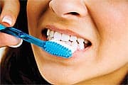
Brush with fluoride toothpaste after eating or drinking. Brush your teeth at least twice a day and ideally after every meal, using fluoride-containing toothpaste. To clean between your teeth, floss or use an interdental cleaner. If you can't brush after eating, at least try to rinse your mouth with water. If you have a young child, ask the dentist how much fluoride toothpaste to put on your child's toothbrush so your child gets the cavity-fighting benefits without getting too much fluoride.
Rinse your mouth. If your dentist feels you have a high risk of developing cavities, he or she may recommend that you use a mouth rinse with fluoride.
Visit your dentist regularly.Get professional teeth cleanings and regular oral exams, which can help prevent problems or spot them early. Your dentist can recommend a schedule that's best for you.
Consider dental sealants.A sealant is a protective plastic coating that's applied to the chewing surface of back teeth, sealing off the grooves and crannies that tend to collect food. The sealant protects tooth enamel from plaque and acid. Sealants can help both children and adults.The Centers for Disease Control and Prevention recommends sealants for all school-age children. Sealants last up to 10 years before they need to be replaced, though they need to be checked regularly to ensure they're still intact.
Drink some tap water. Most public water supplies have added fluoride, which has helped decrease tooth decay significantly. If you drink only bottled water that doesn't contain fluoride, you'll miss out on fluoride benefits.
Avoid frequent snacking and sipping. Whenever you eat or drink beverages other than water, you help your mouth bacteria create acids that can destroy your tooth enamel. If you snack or drink throughout the day, your teeth are under constant attack.
Eat tooth-healthy foods.Some foods and beverages are better for your teeth than others. Avoid foods that get stuck in grooves and pits of your teeth for long periods, such as chips, candy or cookies, or brush soon after eating them. However, foods such as fresh fruits and vegetables increase saliva flow, and unsweetened coffee, tea and sugar-free gum help wash away food particles.
Consider fluoride treatments. Your dentist may recommend periodic fluoride treatments, especially if you aren't getting enough fluoride through fluoridated drinking water and other sources.
Ask about antibacterial treatments. If you're especially vulnerable to tooth decay — for example, because of a medical condition — your dentist may recommend special antibacterial mouth rinses or other treatments to help cut down on harmful bacteria in your mouth.
Most dentists recommend regular checkups to identify cavities and other dental conditions before they cause troubling symptoms and lead to more-serious problems. The sooner you seek care, the better your chances of reversing the earliest stages of tooth decay and preventing its progression. If a cavity is treated before it starts causing pain, you probably won't need extensive treatment.
Treatment of cavities depends on how severe they are and your particular situation. Treatment options include:
Fluoride treatments. If your cavity is just getting started, a fluoride treatment may help restore your tooth's enamel. Professional fluoride treatments contain more fluoride than the amount found in tap water, over-the-counter toothpaste and mouth rinses. Fluoride treatments may be liquid, gel, foam or varnish that's brushed onto your teeth or placed in a small tray that fits over your teeth. Each treatment takes a few minutes.
Fillings. Fillings, sometimes called restorations, are the main treatment option when decay has progressed beyond the earliest enamel-erosion stage. Fillings are made of various materials, such as tooth-colored composite resins, porcelain or combinations of several materials. Silver amalgam fillings contain a variety of materials, including small amounts of mercury.
Crowns. If you have extensive decay or weakened teeth, you may need a crown — a custom-fitted covering that replaces your tooth's entire natural crown. Your dentist will drill away all the decayed area and enough of the rest of your tooth to ensure a good fit. Crowns may be made of gold, porcelain, resin, porcelain fused to metal or other materials.
Root canals. When decay reaches the inner material of your tooth (pulp), you may need a root canal. This is a treatment to repair and save a badly damaged or infected tooth instead of removing it. The diseased tooth pulp is removed. Medication is sometimes put into the root canal to clear any infection. Then the pulp is replaced with a filling.
Tooth extractions. Some teeth become so severely decayed that they can't be restored and must be removed. Having a tooth pulled can leave a gap that allows your other teeth to shift. If possible, consider getting a bridge or a dental implant to replace the missing tooth.
Oral hygiene plays a critical role in whole-body health that is sadly overlooked by most doctors.
As a front-line shield against systemic inflammation, one’s oral status profoundly impacts diseases ranging from type 2 diabetes and cancer to rheumatoid arthritis and atherosclerosis.1-8
Recent scientific studies show that many of the nutrients we now swallow also confer benefits when topically applied in the mouth.9-17
Acting as powerful allies in the fight against periodontal disease, these natural compounds can help safeguard against lethal age-related diseases that emanate from our mouths.
The Gums: An Ideal Incubator for Disease
The oral cavity is a near-perfect breeding ground for microorganisms that lead to decay of gums, teeth, and bone.
Cavities and gum problems that occur in early life are just the beginning. Chronic low-grade inflammation affecting the gums (gingivitis) and inflammation affecting the gums and bones supporting the teeth (periodontitis) has been implicated in the promotion of a variety of insidious systemic disorders, such as coronary heart disease,18 arthritis,6 and even cancer.8,19
Oral inflammation has also been clearly linked to elevated markers of inflammation, such as C-reactive protein.20,21 In response to these warning signs, scientists noted recently, “Evidence for a link between periodontal disease and several systemic diseases is growing rapidly.
Gum Disease and Stroke
Gum disease sets the stage for an increased risk of stroke. A recent review of literature on periodontal disease, published in the Journal of the American Dental Association, concludes that periodontitis among older individuals is associated with an increased risk of developing systemic diseases such as diabetes mellitus, heart attack, and stroke.23 Meanwhile, studies show that efforts to reduce the severity of periodontitis help reduce systemic inflammation and may thereby reduce the risk of cardiovascular events linked with inflammation.
It may arise as a primarylesionoriginating in any of thetissuesin the mouth, bymetastasisfrom a distant site of origin, or by extension from a neighboring anatomic structure, such as thenasal cavity. Alternatively, the oral cancers may originate in any of the tissues of the mouth, and may be of variedhistologictypes:teratoma,adenocarcinomaderived from a major or minorsalivary gland,lymphomafromtonsillaror otherlymphoidtissue, ormelanomafrom the pigment-producing cells of the oral mucosa. There are several types of oral cancers, but around 90% aresquamous cell carcinomas,originating in the tissues that line the mouth and lips. Oral or mouth cancer most commonly involves thetongue. It may also occur on thefloor of the mouth, cheek lining,gingiva(gums), lips, or palate (roof of the mouth). Most oral cancers look very similar under the microscope and are calledsquamous cell carcinoma, but less commonly other types of oral cancer occur, such asKaposi's sarcoma.Oral cancer or mouth cancer, a type of head and neck cancer, is any cancerous tissue growth located in the oral cavity.
Signs and symptoms
On biopsy, the three exophytic masses turned out to be oral carcinomas, while the surrounding hyperkeratotic area showed histologic features of oral lichen planus.
Skin lesion, lump, or ulcer that do not resolve in 14 days located:
On the tongue, lip, or other mouth areas
Usually small
Most often pale colored, may be dark or discolored
Early sign may be a white patch (leukoplakia) or a red patch (erythroplakia) on the soft tissues of the mouth
Usually painless initially
May develop a burning sensation or pain when the tumor is advanced
Behind the wisdom tooth
Even behind the ear
Additional symptoms that may be associated with this disease:
Oncogenes are activated as a result of mutation of the DNA. Risk factors that predispose a person to oral cancer have been identified in epidemiological (epidemiology) studies.
Around 75 percent of oral cancers are linked to modifiable behaviors such as tobacco use and excessive alcohol consumption. Other factors include poor oral hygiene, irritation caused by ill-fitting dentures and other rough surfaces on the teeth, poor nutrition, and some chronic infections caused by bacteria or viruses. If oral cancer is diagnosed in its earliest stages, treatment is generally very effective.
Oral cancer often presents as a non-healing ulcer (shows no sign of healing after 2 weeks). In the US oral cancer accounts for about 8 percent of all malignant growths. Men are affected twice as often as women, particularly men older than
Premalignant lesions
Oral leukoplakia on the buccal mucosa. Overall, leukoplakia carries a risk of transformation to squamous cell carcinoma that ranges from almost 0% to about 20%, which may occur in 1–30 years.[7]
A premalignant (or precancerous) lesion is defined as "a benign, morphologically altered tissue that has a greater than normal risk of malignant transformation." There are several different types of premalignant lesion that occur in the mouth. Some oral cancers begin as white patches (leukoplakia), red patches (erythroplakia) or mixed red and white patches (erythroleukoplakia or "speckled leukoplakia"). Other common premalignant lesions include oral lichen planus (particularly the erosive type), oral submucous fibrosis and actinic cheilitis. In the Indian subcontinent oral submucous fibrosis is very common. This condition is characterized by limited opening of mouth and burning sensation on eating of spicy food. This is a progressive lesion in which the opening of the mouth becomes progressively limited, and later on even normal eating becomes difficult. It occurs almost exclusively in India and Indian communities living abroad. The overall prevalence of oral potentially malignant disorders in the Middle East was 2.8%. Lichen planus/lichenoid lesions were the most common lesions (1.8%) followed by leukoplakias (0.48%), chronic hyperplastic candidiosis (0.38%), and erythroplakia (0.096%). Smoking, alcohol, and age were the main identifiable risk factors.
Tobacco
Oral cancer in a 40-year-old male smoker
In a study of Europeans, smoking and other tobacco use was associated with about 75 percent of oral cancer cases,[10]caused by irritation of the mucous membranes of the mouth from smoke and heat of cigarettes, cigars, and pipes. Tobacco contains over 60 known carcinogens, and the combustion of it, and by-products from this process, is the primary mode of involvement. Use of chewing tobacco or snuff causes irritation from direct contact with the mucous membranes.
Tobacco use in any form by itself, and even more so in combination with heavy alcohol consumption, continues to be an important risk factor for oral cancer. However, due to the current trends in the spread of HPV16, as of early 2011 the virus is now considered the primary causative factor in 63% of newly diagnosed patients.
Alcohol
Some studies in Australia, Brazil and Germany pointed to alcohol-containing mouthwashes as also being etiologic agents in the oral cancer risk family. The claim was that constant exposure to these alcohol-containing rinses, even in the absence of smoking and drinking, leads to significant increases in the development of oral cancer. However, studies conducted in summarize that alcohol-containing mouth rinses are not associated with oral cancer. In a March 2009 brief, the American Dental Association said "the available evidence does not support a connection between oral cancer and alcohol-containing mouthrinse". A 2008 study suggests that acetaldehyde (a breakdown product of alcohol) is implicated in oral cancer, but this study specifically focused on abusers of alcohol and made no reference to mouthwash. Any connection between oral cancer and mouthwash is tenuous without further investigation.
Human papillomavirus
Infection withhuman papillomavirus(HPV), particularly type 16 (there are over 180 types), is a known risk factor and independent causative factor for oral cancer. (Gillison et al. Johns Hopkins) A fast-growing segment of those diagnosed does not present with the historic stereotypical demographics. Historically that has been people over 50, blacks over whites 2 to 1, males over females 3 to 1, and 75% of the time people who have used tobacco products or are heavy users of alcohol. This new and rapidly growing sub population between 30 and 50 years old,[17]is predominantly nonsmoking, white, and males slightly outnumber females. Recent research from multiple peer-reviewed journal articles indicates that HPV16 is the primary risk factor in this new population of oral cancer victims. HPV16 (along with HPV18) is the same virus responsible for the vast majority of allcervical cancersand is the most common sexually transmitted infection in the US. Oral cancer in this group tends to favor the tonsil and tonsillar pillars, base of the tongue, and theoropharynx. Recent data suggest that individuals that come to the disease from this particular etiology have a significant survival advantage, as the disease responds better to radiation treatments than tobacco etiology disease.
Hematopoietic stem cell transplantation
Patients after hematopoietic stem cell transplantation (HSCT) are at a higher risk for oral squamous cell carcinoma. Post-HSCT oral cancer may have more aggressive behavior with poorer prognosis, when compared to oral cancer in non-HSCT patients.[18] This effect is supposed to be owing to the continuous lifelong immune suppression and chronic oral graft-versus-host disease.
Diagnosis
Histopathologic appearance of a well differentiated squamous cell carcinoma specimen. Hematoxylin-eosin stain
Early diagnosis of oral cancer patients would decrease mortality and help to improve treatment. Oral surgeons and dentists are the early diagnosers that diagnose these patients in early stages. Health providers, dentists, and oral surgeons shall have high knowledge and awareness that would help them to provide better diagnosis for oral cancer patients. An examination of the mouth by the health care provider, dentist, oral surgeons shows a visible and/or palpable (can be felt)lesion of the lip, tongue, or other mouth area. The lateral/ventral sides of the tongue are the most common sites for intraoral SCC. As the tumor enlarges, it may become an ulcer and bleed. Speech/talking difficulties, chewing problems, or swallowing difficulties may develop. A feeding tube is often necessary to maintain adequate nutrition. This can sometimes become permanent as eating difficulties can include the inability to swallow even a sip of water. The doctor can order some special investigations which may include a chest x-ray, CT or MRI scans, and tissue biopsy.
There are a variety of screening devices that may assist dentists in detecting oral cancer, including the Velscope, Vizilite Plus and the identafi 3000. There is no evidence that routine use of these devices in general dental practice saves lives.[19] However, there are compelling reasons to be concerned about the risk of harm this device may cause if routinely used in general practice. Such harms include false positives, unnecessary surgical biopsies and a financial burden on the patient. While a dentist, physician or other health professional may suspect a particular lesion is malignant, there is no way to tell by looking alone - since benign and malignant lesions may look identical to the eye. A non-invasive brush biopsy (BrushTest) can be performed to rule out the presence of dysplasia (pre-cancer) and cancer on areas of the mouth that exhibit an unexplained color variation or lesion. The only definitive method for determining if cancerous or precancerous cells are present is through biopsy and microscopic evaluation of the cells in the removed sample. A tissue biopsy, whether of the tongue or other oral tissues and microscopic examination of the lesion confirm the diagnosis of oral cancer or precancer. There are six common species of bacteria found at significantly higher levels in the saliva of patients with oral squamous cell carcinoma (OSCC) than in saliva of oral-free cancer individuals. Three of the six, C. gingivalis, P. melaninogenica, and S. mitis, can be used as a diagnostic tool to predict more than 80% of oral cancers.
Management
Surgical excision (removal) of the tumor is usually recommended if the tumor is small enough, and if surgery is likely to result in a functionally satisfactory result. Radiation therapy with or withoutchemotherapy is often used in conjunction with surgery, or as the definitive radical treatment, especially if the tumour is inoperable. Surgeries for oral cancers include:
Mandibulectomy (removal of the mandible or lower jaw or part of it)
Glossectomy (tongue removal, can be total, hemi or partial). When glossectomy is performed for smaller tumors (< 4 cm), the adequacy of resection (margin status) is best assessed from the resected specimen itself. The status of the margin (positive/tumor cut through versus negative/clear margin) obtained from the glossectomy specimen appears to be of prognostic value, while the status of the margin sampled from the post-glossectomy defect is not. The method of margin sampling appears to correlate with local recurrence: preference for tumor bed/defect margins may be associated with worse local control.[21][22]
Combinational, e.g. glossectomy and laryngectomy done together
Feeding tube to sustain nutrition
Owing to the vital nature of the structures in the head and neck area, surgery for larger cancers is technically demanding. Reconstructive surgery may be required to give an acceptable cosmetic and functional result. Bone grafts and surgical flaps such as the radial forearm flap are used to help rebuild the structures removed during excision of the cancer. An oral prosthesis may also be required. Most oral cancer patients depend on a feeding tube for their hydration and nutrition. Some will also get a port for the chemo to be delivered. Many oral cancer patients are disfigured and suffer from many long term after effects. The after effects often include fatigue, speech problems, trouble maintaining weight, thyroid issues, swallowing difficulties, inability to swallow, memory loss, weakness, dizziness, high frequency hearing loss and sinus damage.
Survival rates for oral cancer depend on the precise site, and the stage of the cancer at diagnosis. Overall, 2011 data from the SEER database shows that survival is around 57% at five years when all stages of initial diagnosis, all genders, all ethnicities, all age groups, and all treatment modalities are considered. Survival rates for stage 1 cancers are approximately 90%, hence the emphasis on early detection to increase survival outcome for patients. Similar survival rates are reported from other countries such as Germany.
Chemotherapy is useful in oral cancers when used in combination with other treatment modalities such as radiation therapy. It is not used alone as a monotherapy. When cure is unlikely it can also be used to extend life and can be considered palliative but not curative care. Biological agents, such as Cetuximab have recently been shown to be effective in the treatment of squamous cell head and neck cancers, and are likely to have an increasing role in the future management of this condition when used in conjunction with other established treatment modalities.
Treatment of oral cancer will usually be by a multidisciplinary team, with treatment professionals from the realms of radiation, surgery, chemotherapy, nutrition, dental professionals, and even psychology all possibly involved with diagnosis, treatment, rehabilitation, and patient care.
Prognosis
Postoperative disfigurement of the face, head and neck
Complications of radiation therapy, including dry mouth and difficulty swallowing
Prognosis depends on stage and overall health. Grading of the invasive front of the tumor is a very important prognostic parameter
Optimizing oral health represents a crucial disease-prevention strategy.
Poor oral health, particularly periodontitis, can contribute to a wide range of serious diseases ranging from rheumatoid arthritis to stroke and even cancer.
Bacteria thriving in the oral cavity contribute to inflammation, which can have detrimental effects throughout the body and may particularly increase the risk of atherosclerosis. Oral bacteria have even been found living inside atherosclerotic plaques.
Natural agents such as green tea, aloe vera, lactoferrin, xylitol, folic acid, and hydrogen peroxide can help optimize oral health by targeting plaque-causing bacteria and supporting gum health.
Coenzyme Q10 (CoQ10) shows particular promise in offsetting the inflammation that accompanies gum disease. CoQ10 offers benefits both when applied topically to the gums and when consumed as a dietary supplement. The benefits of supplemental CoQ10 begin at a daily dose of 50 mg.
Pomegranate helps preserve periodontal health by preventing the adherence of plaque-inducing bacteria to the teeth, by directly killing oral microorganisms, and by quieting inflammation in the gums.
Vitamins C and D may also promote healthy teeth and gums.
A two-pronged approach that includes brushing the teeth and rinsing the mouth with natural bioactive ingredients provides the foundation of oral health—a cornerstone of whole-body wellness.
Gum Disease and Obesity
Some researchers are now suggesting that perio-dontitis may contribute to obesity by elevating C-reactive protein, which then acts as a potent inducer of inflammatory cytokines and hormones secreted by adipose tissue.26-29
Scientists have found that elevated C-reactive protein causes fat cells (adipocytes) to store more fat and burn less energy. Indeed, evidence is accumulating that there is a link between obesity, type 2 diabetes, and periodontitis. As one research team noted recently, “Obesity is a significant predictor of periodontal disease and insulin resistance appears to mediate this relationship.”28 A University of Mississippi study found, “…significant correlations between body composition and periodontal disease,” and noted this finding “strengthened arguments that periodontal disease and certain obesity-related systemic illnesses are related…”29
Periodontal Disease Linked With Cancer
The link between oral health and cancer remains somewhat controversial, largely because this information is so new. But a recently published study by researchers at the Imperial College of London and Harvard School of Public Health has shed new light on the matter. By carefully eliminating potential confounding factors, such as a patient’s history of cigarette smoking, these scientists sought to identify any statistically significant associations between oral health and the incidence of cancer. Their conclusion is chilling. “Periodontal disease was associated with a small, but significant, increase in overall cancer risk, which persisted in never-smokers,” write the collaborators, in the medical journal Lancet Oncology.19 This conclusion has profound implications. The fact that it arises from data gathered from more than 48,000 men over the course of approximately 18 years lends additional gravity to the findings.
The research team also found significant associations among oral health status and lung, kidney, and pancreatic cancers, as well as cancers of the blood. The investigators note that their results need independent confirmation, but they offer this speculation regarding the implications of the findings: “…periodontal disease might be a marker of a susceptible immune system or might directly affect cancer risk.”19 In either case, periodontal disease takes on new significance, and appears to pose more of a threat to health than has previously been recognized.
Furthermore, a recent study by researchers at the Harvard School of Public Health tentatively concludes that periodontitis is associated with an increased risk of one of the most deadly cancers. “Compared with no periodontal disease, history of periodontal disease was associated with increased pancreatic cancer risk,” write the Harvard researchers, in the Journal of the National Cancer Institute.30
The American Dental Association agrees that “oral health is important for overall health” and indicates that salivary diagnosis may offer a key tool in health assessment. “A wide range of proteins, nucleic acids, hormones, pharmaceuticals, and pathogens can be measured in saliva, making it an excellent candidate for rapid detection and screening of biomarkers for conditions like caries, periodontal disease, osteoporosis, infectious diseases, and cancer,” it says.31
ABOUT PERIODONTAL DISEASE
Bacteria and other microorganisms are the underlying cause of tooth decay. Bacteria break down compounds from food called fermentable carbohydrates (e.g. sucrose), producing lactic acid and other organic acids as a byproduct. These acids promote enamel and dentin demineralization. This softening of the enamel then leads to the development of dental caries (cavities).
Although bacteria naturally co-exist with us, under certain conditions they form a biofilm. A biofilm is a sort of living carpet composed of various bacteria and even fungi.12 The microorganisms excrete a kind of glue, firmly anchoring themselves to the enamel surface of the tooth. Biofilm formation, and especially biofilm attachment, is at the root of dental disease.57 Plaque is the common term for this living aggregation of various bacteria and fungi. Over time, plaque hardens and takes on various minerals. At this stage, the coating is called tartar. It is this hard coating that dental hygienists work to scrape away in the dentist’s office.
Gingivitis occurs when dental plaque stimulates an immune response in the soft tissues surrounding the teeth. The gums become inflamed and irritated, appearing swollen and red and bleeding easily. If gingivitis is left untreated, it may progress to periodontitis, a condition in which Gram-negative bacteria destroy the supportive structures of the teeth. Periodontitis may ultimately lead to tooth loss.
Avoiding the buildup of plaque is the reason dentists encourage us to brush our teeth and to floss regularly. Brushing mechanically breaks up the film to some extent and rinsing helps remove fermentable sugars. But on the biochemical level, there is more that can be done to fight what is, after all, a biological enemy. This is where natural bioactive agents that target plaque microorganisms and promote gum healing come in.
Botanical and Nutritional Agents Show Promise in Oral Hygiene
Given the potentially lethal risks of poor dental hygiene, it makes sense to utilize all the science available to prevent even the smallest problems in the mouth.
Several nutrients have shown very favorable effects when used as part of an oral hygiene program. Among these are coenzyme Q10 (CoQ10), green tea, aloe vera, and pomegranate. These claims have been verified by published research.9-11,32-35 Other beneficial ingredients for healthy teeth and gums include xylitol, lactoferrin, and folic acid.12,13,15,17
Multi-Faceted Benefits of Green Tea
Green tea is well known for its beneficial effects throughout the body, but it is also effective in the fight against dental caries and oral disease. Studies have shown that green tea catechins exert direct antibacterial activity against Streptococcus mutans, one of the key microorganisms responsible for tooth decay. Green tea also helps prevent bacteria from sticking to teeth, by inhibiting a bacterial enzyme involved in this process. It also inhibits production of amylase, an enzyme used by bacteria to break starches down into sugars, which bacteria use to fuel their own growth.34,35
Furthermore, Asian researchers showed recently that green tea reduces the invasiveness of oral cancer and decreases the production of a protein associated with oral cancer proliferation.36,37 Additionally, American researchers report that green tea arrests the growth and causes self-destruction (apoptosis) of oral carcinoma cells in the laboratory.38
In Japan, researchers conducted a study in which green tea was applied to the teeth of subjects with periodontal disease for eight weeks. Symptoms of periodontitis improved in subjects receiving green tea catechins and there was objective evidence that green tea killed a significant proportion of the bacteria causing periodontitis in these test subjects.39
Fight Oral Disease
Best known as a potent cardioprotective nutrient, CoQ10 has also been shown to improve symptoms of periodontitis when applied topically in the oral cavity.9,32,33 Japanese researchers conducted a placebo-controlled clinical trial in men with established periodontitis. After nine weeks of CoQ10 application, investigators found evidence of “significant improvements” in periodontal status, which were not seen in control subjects.9
An early study on CoQ10’s effectiveness against periodontitis impressed the study’s authors so much, they wrote, “Healing was so excellent five to seven days’ post-biopsy that the biopsy sites were difficult to locate. The healing was viewed as extraordinarily effective.”40 It has been suggested that CoQ10 benefits oral health by reducing the oxidative stress associated with low-grade inflammation of gums and bone.
Good oral and dental hygiene can help you avoid cavities and tooth decay. Below are some tips to help prevent cavities. Ask your dentist which tips are best for you.
Brush with fluoride toothpaste after eating or drinking. Brush your teeth at least twice a day and ideally after every meal, using fluoride-containing toothpaste. To clean between your teeth, floss or use an interdental cleaner. If you can't brush after eating, at least try to rinse your mouth with water. If you have a young child, ask the dentist how much fluoride toothpaste to put on your child's toothbrush so your child gets the cavity-fighting benefits without getting too much fluoride.
Rinse your mouth. If your dentist feels you have a high risk of developing cavities, he or she may recommend that you use a mouth rinse with fluoride.
Visit your dentist regularly.Get professional teeth cleanings and regular oral exams, which can help prevent problems or spot them early. Your dentist can recommend a schedule that's best for you.
Consider dental sealants.A sealant is a protective plastic coating that's applied to the chewing surface of back teeth, sealing off the grooves and crannies that tend to collect food. The sealant protects tooth enamel from plaque and acid. Sealants can help both children and adults.The Centers for Disease Control and Prevention recommends sealants for all school-age children. Sealants last up to 10 years before they need to be replaced, though they need to be checked regularly to ensure they're still intact.
Drink some tap water. Most public water supplies have added fluoride, which has helped decrease tooth decay significantly. If you drink only bottled water that doesn't contain fluoride, you'll miss out on fluoride benefits.
Avoid frequent snacking and sipping. Whenever you eat or drink beverages other than water, you help your mouth bacteria create acids that can destroy your tooth enamel. If you snack or drink throughout the day, your teeth are under constant attack.
Eat tooth-healthy foods.Some foods and beverages are better for your teeth than others. Avoid foods that get stuck in grooves and pits of your teeth for long periods, such as chips, candy or cookies, or brush soon after eating them. However, foods such as fresh fruits and vegetables increase saliva flow, and unsweetened coffee, tea and sugar-free gum help wash away food particles.
Consider fluoride treatments. Your dentist may recommend periodic fluoride treatments, especially if you aren't getting enough fluoride through fluoridated drinking water and other sources.
Ask about antibacterial treatments. If you're especially vulnerable to tooth decay — for example, because of a medical condition — your dentist may recommend special antibacterial mouth rinses or other treatments to help cut down on harmful bacteria in your mouth.
Most dentists recommend regular checkups to identify cavities and other dental conditions before they cause troubling symptoms and lead to more-serious problems. The sooner you seek care, the better your chances of reversing the earliest stages of tooth decay and preventing its progression. If a cavity is treated before it starts causing pain, you probably won't need extensive treatment.
Treatment of cavities depends on how severe they are and your particular situation. Treatment options include:
Fluoride treatments. If your cavity is just getting started, a fluoride treatment may help restore your tooth's enamel. Professional fluoride treatments contain more fluoride than the amount found in tap water, over-the-counter toothpaste and mouth rinses. Fluoride treatments may be liquid, gel, foam or varnish that's brushed onto your teeth or placed in a small tray that fits over your teeth. Each treatment takes a few minutes.
Fillings. Fillings, sometimes called restorations, are the main treatment option when decay has progressed beyond the earliest enamel-erosion stage. Fillings are made of various materials, such as tooth-colored composite resins, porcelain or combinations of several materials. Silver amalgam fillings contain a variety of materials, including small amounts of mercury.
Crowns. If you have extensive decay or weakened teeth, you may need a crown — a custom-fitted covering that replaces your tooth's entire natural crown. Your dentist will drill away all the decayed area and enough of the rest of your tooth to ensure a good fit. Crowns may be made of gold, porcelain, resin, porcelain fused to metal or other materials.
Root canals. When decay reaches the inner material of your tooth (pulp), you may need a root canal. This is a treatment to repair and save a badly damaged or infected tooth instead of removing it. The diseased tooth pulp is removed. Medication is sometimes put into the root canal to clear any infection. Then the pulp is replaced with a filling.
Tooth extractions. Some teeth become so severely decayed that they can't be restored and must be removed. Having a tooth pulled can leave a gap that allows your other teeth to shift. If possible, consider getting a bridge or a dental implant to replace the missing tooth.


_squamous_cell_carcinoma_histopathology.jpg)