Disease Topics:Burns

Burns are injuries to tissues caused by heat, friction, electricity, radiation, or chemicals.

Description

Burns are characterized by degree, based on the severity of the tissue damage. A first-degreeburn causes redness and swelling in the outermost layers of skin (epidermis). A second-degreeburn involves redness, swelling and blistering, and the damage may extend beneath theepidermis to deeper layers of skin (dermis). A third-degree burn, also called a full-thickness burn,destroys the entire depth of skin, causing significant scarring. Damage also may extend to theunderlying fat, muscle, or bone.
The severity of the burn is also judged by the amount of body surface area (BSA) involved. Healthcare workers use the "rule of nines" to determine the percentage of BSA affected in patientsmore than 9 years old: each arm with its hand is 9% of BSA; each leg with its foot is 18%; thefront of the torso is 18%; the back of the torso, including the buttocks, is 18%; the head andneck are 9%; and the genital area (perineum) is 1%. This rule cannot be applied to a youngchild's body proportions, so BSA is estimated using the palm of the patient's hand as a measureof 1% area.
The severity of the burn will determine not only the type of treatment, but also where the burnpatient should receive treatment. Minor burns may be treated at home or in a doctor's office.These are defined as first- or second-degree burns covering less than 15% of an adult's body orless than 10% of a child's body, or a third-degree burn on less than 2% BSA. Moderate burnsshould be treated at a hospital. These are defined as first- or second-degree burns covering15%-25% of an adult's body or 10%-20% of a child's body, or a third-degree burn on 2%-10%BSA. Critical, or major, burns are the most serious and should be treated in a specialized burnunit of a hospital. These are defined as first- or second-degree burns covering more than 25% ofan adult's body or more than 20% of a child's body, or a third-degree burn on more than 10%BSA. In addition, burns involving the hands, feet, face, eyes, ears, or genitals are consideredcritical. Other factors influence the level of treatment needed, including associated injuries suchas bone fractures and smoke inhalationpresence of a chronic disease, or a history of beingabused. Also, children and the elderly are more vulnerable to complications from burn injuriesand require more intensive care.

Causes and symptoms

Burns may be caused by even a brief encounter with heat greater than 120°F (49°C). The sourceof this heat may be the sun (causing a sunburn), hot liquids, steam, fire, electricity, friction(causing rug burns and rope burns), and chemicals (causing a caustic burn upon contact).
Signs of a burn are localized redness, swelling, and pain. A severe burn will also blister. Theskin may also peel, appear white or charred, and feel numb. A burn may trigger a headacheand feverExtensive burns may induce shockthe symptoms of which are faintness, weakness,rapid pulse and breathing, pale and clammy skin, and bluish lips and fingernails.

Diagnosis

physician will diagnose a burn based upon visual examination, and will also ask the patient orfamily members questions to determine the best treatment. He or she may also check forsmoke inhalation, carbon monoxide poisoningcyanide poisoningother event-relatedtrauma, or, if suspected, further evidence of child abuse.


burn

 (bern),
1. sensation of pain caused by excessive heat, or similar pain from any cause.
2. lesion caused by heat or any cauterizing agent, including friction, caustic agents,electricity, or electromagnetic energy; types of burns resulting from different agents arerelatively specific and diagnostic. The division of burns into three levels (superficial, partialthickness, full-thickness, [q.v.]) reflects the severity of skin damage (erythema, blisters,charring, respectively).
[A.S. baernan]

burn

 (burninjury to tissues caused by the contact with heat, flame, chemicals, electricity, orradiation. First degree burns show redness; second degree burns show vesication; third degreeburns show necrosis through the entire skin. Burns of the first and second degree are partial-thickness burns, those of the third degree are full-thickness burns.
first-degree burn  a burn that affects the epidermis only, causing erythema without blistering.
fourth-degree burn  a burn that extends deeply into the subcutaneous tissue; it may involvemuscle, fascia, or bone.
full-thickness burn  third-degree b.
partial-thickness burn  second-degree b.
second-degree burn  a burn that affects the epidermis and the dermisclassified assuperficial (involving the epidermis and the papillary dermis) or deep (extending into the reticulardermis). Called also partial thickness b.
third-degree burn  a burn that destroys both the epidermis and the dermisoften alsoinvolving the subcutaneous tissue. Called also full-thickness b.burn
Etymology: AS, baernan
any injury to tissues of the body caused by hot objects or flames, electricity, chemicals,radiation, or gases in which the extent of the injury is determined by the nature of the agent,length of time exposed, body part involved, and depth of burn. The treatment of burns includespain relief, careful asepsis, prevention of infection, regulation of body temperature, maintenanceof the balance in the body of fluids and electrolytes, and good nutrition. First priority with burns ofthe airway is airway control. Severe burns of any origin may cause shock, which is treatedbefore the wound. Burns are sometimes classified as first, second, third, and fourth degree.First-degree burns involve only a superficial layer of epidermal cells. Second-degree burns maybe divided into superficial partial-thickness and deep partial-thickness wounds. Damage insecond-degree burns extends through the epidermis to the dermis but is usually not sufficient toprevent skin regeneration. In third-degree burns the entire thickness of the epidermis and dermisis destroyed. Fourth-degree burns are full-thickness injuries that penetrate the subcutaneoustissue, muscle, and periosteum or bone. See also chemical burnelectrocutionthermal burn.
enlarge picture
Deep partial-thicknesssecond-degree burnburn
 (burn)
Enlarge picture
SECOND DEGREE BURN
Enlarge picture
BURNS
Enlarge picture
BURNS
Enlarge picture
BURNS
Tissue injury resulting from excessive exposureto thermal, chemical, electrical, or radioactiveagents. The effects may be local, resulting incell injury or death, or both local and 

acid burn

burn caused by exposure to corrosive acids such as sulfuric, hydrochloric, and nitric.

actinic burns

Burns caused by ultraviolet or sun rays. Treatment is the same as for dry heat burns.

burn of aerodigestive tract

Necrosis of the oral mucosa, trachea, or esophagus due to the ingestion of caustic substances.After an assessment of the patient's airway, breathing, and circulation, the medical teamdetermines the severity of the exposure by physical examination or laryngoscopy. Some patientsmay require hospitalization for local care and the administration of intravenous steroids, histamineantagonists, and antibiotics. Late complications may include strictures of the affected internalorgans.

alkali burn


A burn caused by caustic alkalies such as lye, caustic potash (potassium hydroxide), and causticsoda (sodium hydroxide), and marked by a painful skin lesion, often associated with gelatinizationof tissue.


Treatment

Burn treatment consists of relieving pain, preventing infection, and maintaining body fluids,electrolytes, and calorie intake while the body heals. Treatment of chemical or electrical burns isslightly different from the treatment of thermal burns but the objectives are the same.

Thermal burn treatment

The first act of thermal burn treatment is to stop the burning process. This may be accomplishedby letting cool water run over the burned area or by soaking it in cool (not cold) water. Ice shouldnever be applied to the burn. Cool (not cold) wet compresses may provide some pain relief whenapplied to small areas of first- and second-degree burns. Butter, shortening, or similar salveshould never be applied to the burn since it prevents heat from escaping and drives the burningprocess deeper into the skin.

Treatment

Burn treatment consists of relieving pain, preventing infection, and maintaining body fluids,electrolytes, and calorie intake while the body heals. Treatment of chemical or electrical burns isslightly different from the treatment of thermal burns but the objectives are the same.

Thermal burn treatment


The first act of thermal burn treatment is to stop the burning process. This may be accomplishedby letting cool water run over the burned area or by soaking it in cool (not cold) water. Ice shouldnever be applied to the burn. Cool (not cold) wet compresses may provide some pain relief whenapplied to small areas of first- and second-degree burns. Butter, shortening, or similar salveshould never be applied to the burn since it prevents heat from escaping and drives the burningprocess deeper into the skin.


Treatment

The first responsibility in the care of the burnpatient is to assess the patency of the airwayand to ensure that breathing is unimpaired. Ifsmoke inhalation or airway injury is suspected,intubation should be performed before edemamakes this impossible. Airway injury is mostlikely to occur after facial burns or smokeinhalation in closed spaces. A cough productiveof soot or charred material increases thelikelihood of inhalational injury.

The second task in burn care is to ensurecardiac output and tissue perfusion. Volumeresuscitation with crystalloid is given perstandard protocols; at the same time, urinaryoutput, blood pressure and pulse, body weights,and renal function are closely monitored toensure adequate hydration.

The immediate care of the burn itself involves theremoval of any overlying clothing and jewelry andthe irrigation of the affected tissues with coolwater, taking care to avoid excessively coolingthe body. To help prevent hypothermia andinfection, cover the burn wounds with steriledressings if available, or a clean sheet,separating burn wound surfaces. Gentle tissuedébridement should be followed by application ofnonadherent dressings, skin substitutes, topicalantiseptics, or autografts, as dictated bycircumstances. Tetanus prophylaxis is routinelygiven, usually with both tetanus toxoid andtetanus immune globulin.

In specific circumstances, additionalinterventions such as hyperbaric oxygen therapyfor carbon monoxide intoxication, escharotomyfor circumferential burns, antibiotic therapy forinfections, pressor support for hypotension, ornutritional support may be needed.

Patients with large or complex burn injuriesshould be transferred to regional burn centers orto the care of surgeons with special interest inburn management.

Patient care

During rehabilitation, individually fitted elasticgarments are applied to prevent hypertrophicscar formation, and joints are exercised topromote a full range of motion. The patient isencouraged to increase activity tolerance, obtainadequate rest, strive for physical and emotionalindependence, and resume vocational and socialfunctioning. Referrals for occupational therapy,psychological counseling, support groups, orsocial services are often necessary.Reconstructive and cosmetic surgery may berequired. Support groups and services areavailable to assist the patient with lifeadjustments.

Patients' previous psychological states maypredispose them to injury and may have anadverse effect on recovery. Patients with burninjuries demonstrate a wide range of emotionalresponses including anger, frustration, irritability,and psychological states (delirium, anxiety,depression, and grief). Posttraumatic stressdisorder (PTSD) may occur after a burn injury.Often, the PTSD patient will need help fromprimary or specialized care providers to recoverpsychologically. Explain patient needs and careconcerns to family to help alleviate their caresand concerns (and varied psychologicalresponses). Involve them with you in patient careas permissible. Family members should beencouraged to sit with the patient, and to touch,speak to, read to, and otherwise communicatewith the patient. Providing patients with a senseof purpose will help to alleviate feelings ofhelplessness and will provide both patient andfamily with more comfortable and comfortingmemories.

The provision of optimal nutrition to burn patientsis an important component of recovery. Becauseof protein losses, the total protein consumed bya burn patient should be at least 2.5 g/kg ofbody weight daily. Total caloric needs mayexceed 30 kcal/kd/daily. The risk of infectionsmay be reduced by the provision of dietarysupplements, esp. arginine and glutamine.

Emergency Care


The burn is irrigated with large volumes of water and dressed.

Treatment

Loose dirt is carefully brushed away and the area is cleansed with soap and water. An antisepticsolution or ointment is applied and covered with a dressing. Tetanus toxoid or antitoxin is given ifrequired. A brush burn is also informally called a “road rash” as in the case of a motorcyclist whoslid across the pavement.


chemical burn

Tissue destruction caused by corrosive or irritating chemicals such as strong acids or bases,phenols, pesticides, disinfectants, fertilizers, or chemical warfare agents.

Treatment

Irrigate with large quantities of water.

electric burn

Tissue destruction caused by the passage of electrical current through the body, usually as a resultof industrial accidents or lightning exposures. Entry and exit wounds are usually present; significantinternal organ damage may be found along the path of the current through the body.

burn of eye

burn of the eyeball due to contact with chemical, thermal, electrical, or radioactive agents.

Emergency Care

The eye should be washed immediately with the nearest available supply of water, even if it is notsterile. Irrigation may need to be continued for hours if burn is due to lye. Care must be taken toprevent runoff from draining into the uninjured eye.

fireworks burn

Injury from explosives; usually a burn, often with embedded foreign bodies and a high incidence ofinfection and tetanus, which should be prevented by meticulous care of injury and use of antitetanustoxoid and immune globulin.

flash burn

burn resulting from an explosive blast such as occurs from ignition of highly inflammable fluids, orin war from a high-explosive shell or a nuclear blast.

gunpowder burn

burn resulting from exploding gunpowder, usually at very close range. It is often followed bytetanus, which should be prevented by administration of antitetanus toxoid and immune globulin andmeticulous care of the injury area.

inhalation burn

Inhalation injury.

radiation burn

burn resulting from overexposure to radiant energy as from x-rays, radium or other radioactiveelements, sunlight, or nuclear blast.

respiratory burn

burn to the components of the respiratory system usually caused by inhaling superheated gases.See: inhalation injury
Enlarge picture
BLISTERED PARTIAL THICKNESS THERMALBURN

thermal burn

burn resulting from contact with fire, hotobjects, or fluids.

See: illustration


x-ray burn

Complications

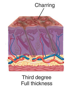
Sloughing of skin, gangrene, scarring,erysipelas, nephritis, pneumonia, immunesystem impairment, or intestinal disturbancesare possible complications. Shock and infectionmust always be anticipated with higher-degreeor larger burns. The risk of complication isgreatest when more than 25% of the bodysurface is burned.

Etiology

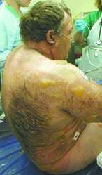
Burns may result from ultraviolet radiation,bursts of steam, heated liquids and metals,chemical fires, electrocution, or direct contactwith flame or flammable clothing.
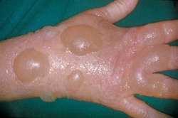
Precautions

person in burning clothing should never beallowed to run. The individual should lie downand roll. A rug, blanket, or anything within reachcan be used to smother the flames. Care mustbe taken so that the individual does not inhalethe smoke. The clothing should be cut offcarefully so that the skin is not pulled away.Synthetic fabrics that have melted into the burnwound are best removed later in the emergencydepartment or burn center. Jewelry should beremoved even if not near the burn wounds due toconcerns for fluid shifts and swelling. Blistersshould not be opened, as this increases thechance for infection. Patients with large burnareas or third- and fourth-degree burns mustreceive appropriate tetanus prophylaxis.

Note

In severe, widespread burns, the patient must betransferred to a burn center as soon as ispractical.